Jump to content
In this section we have spot diagnoses posted on a daily basis since June 2010, now over 4000! You can review the archived cases and read the suggested diagnoses by users and the final comment by the contributors.
Case are uploaded each week day by 10 am UK time with the correct diagnosis will generally be posted at 8 pm UK time. Why not view the most recent spot diagnosis and proffer a diagnosis?

Case Number : Case 920 - 31st December Posted By: Guest

Please read the clinical history and view the images by clicking on them before you proffer your diagnosis.
Submitted Date :
   (0 reviews)

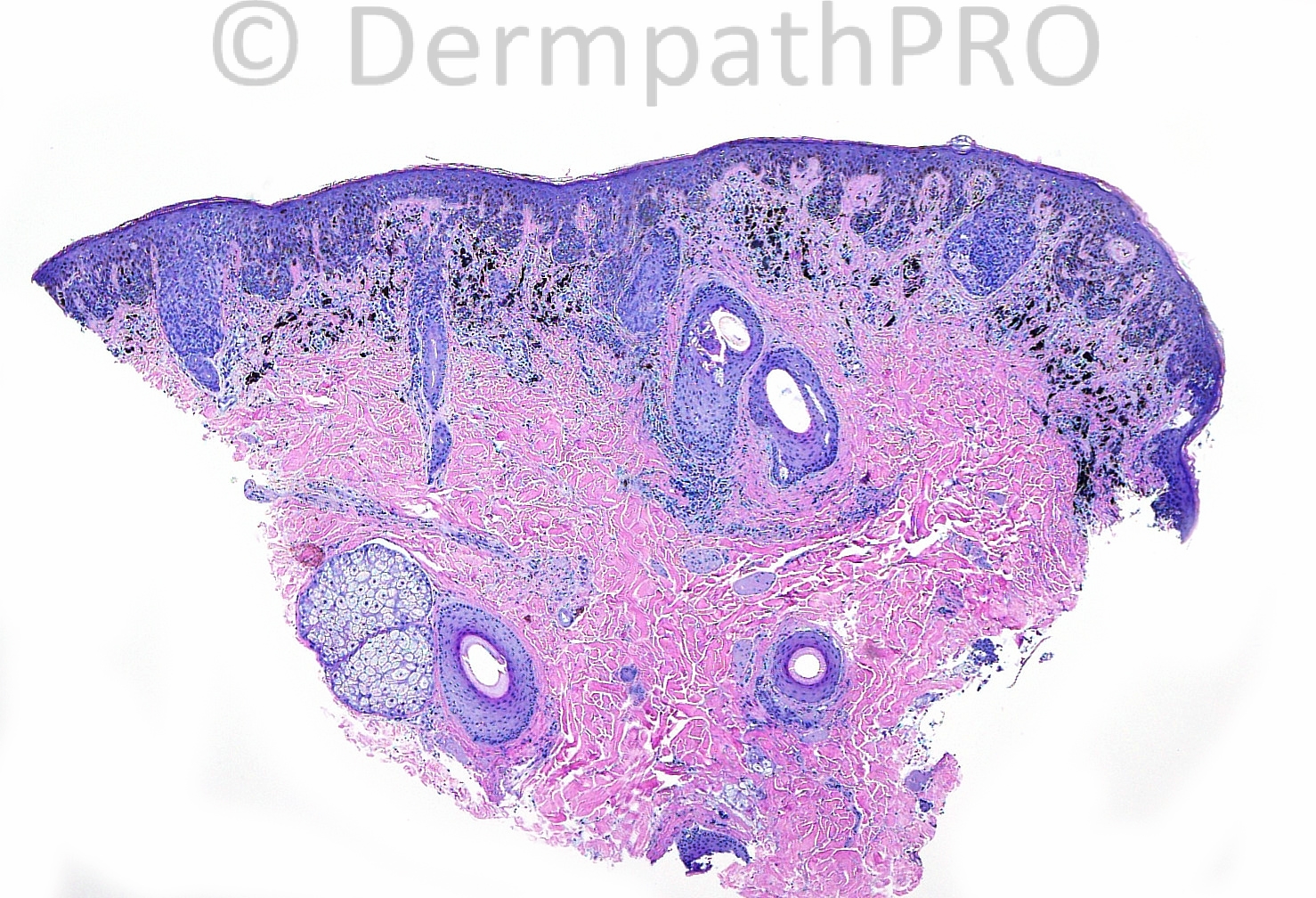
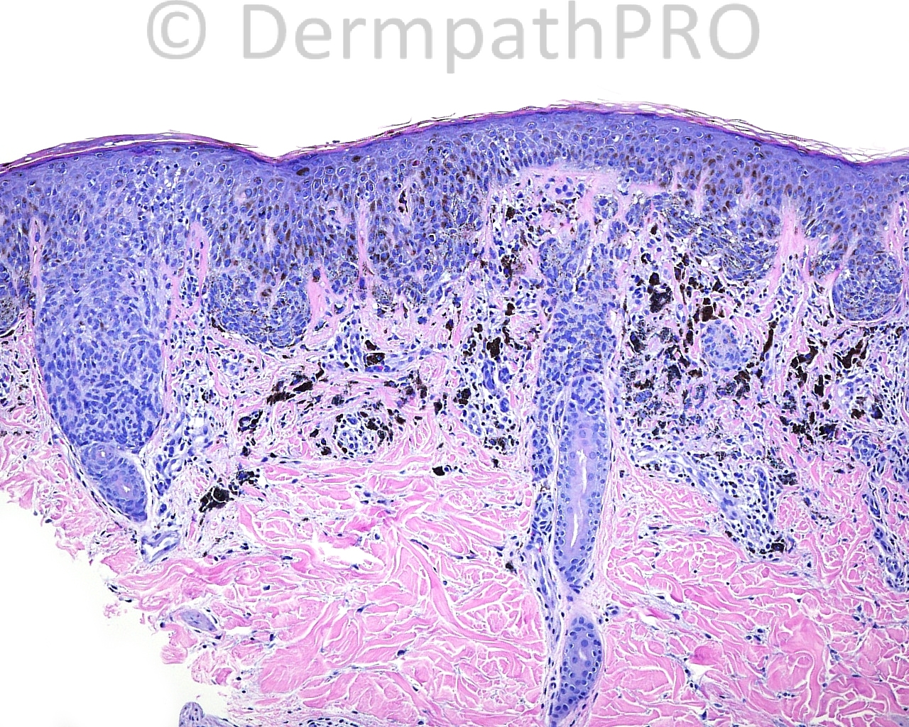
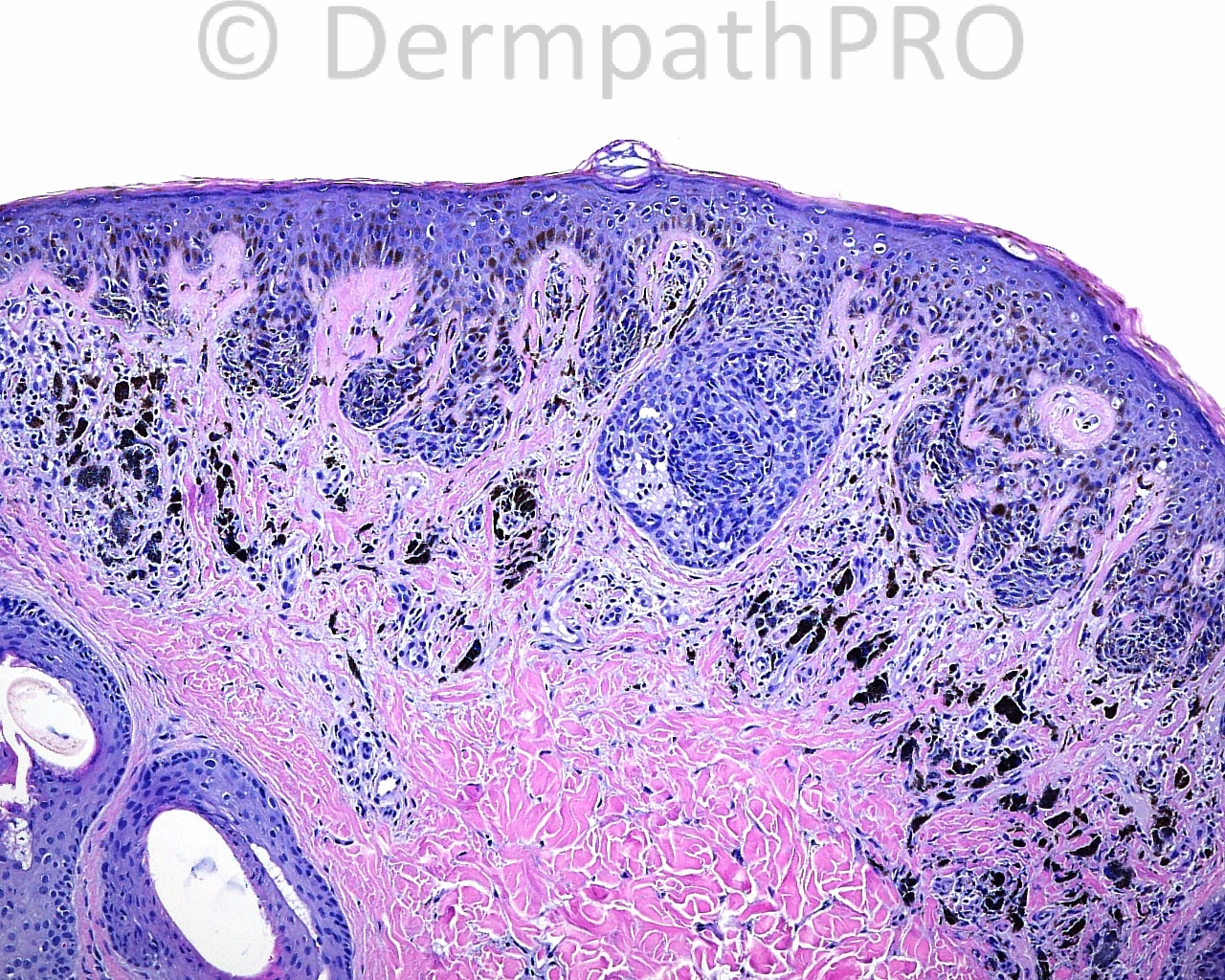
The patient is a 14 year old boy with a punch biopsy taken from the right temporal scalp.

Case posted by Dr. Mark Hurt


  Report Record

User Feedback


Guest Romualdo

Posted

I think this is just an intradermal melanocytic nevus from one of the special sites, to know, the scalp.

Share this comment


Link to comment
share_externally

Sasi Attili

Posted

I would go for a compound naevus with architectural atypia in junctional nests- benign. Complete excision however advised if clinical suspicion persists.

Share this comment


Link to comment
share_externally

Robledo F. Rocha

Posted

Compound melanocytic nevus of so-called special site with secondary trichostasis spinulosa.

Share this comment


Link to comment
share_externally

Eman El-Nabarawy

Posted

Compound melanocytic nevus of special site (scalp).

Share this comment


Link to comment
share_externally

Guest Amira Tawdy

Posted

Atypical pigmented spindle cell nevus ( vertically oriented nests of spindled shaped cells, the nests are irregular in size, few are confluent, fusion of the rete riges in some foci, many dermal melanophages)

Share this comment


Link to comment
share_externally

Dr. Richard Carr

Posted

Although I am not overly worried by the appearances here in practice I would be very reluctant to give anything more than a descriptive report (e.g. dark, junctionally active melanocytic lesion, relatively bland but further comment is limited by lack of clinical information). In my view punch biopsy of a pigmented lesion in the absence of any useful clinical information are the perfect scenario for litigation!

Share this comment


Link to comment
share_externally

Guest Romualdo

Posted

I meant to say "compound", not "intradermal" melanocytic nevus from one of the special sites.

Share this comment


Link to comment
share_externally

Dr. Mona Abdel-Halim

Posted

Agree with Dr Carr... I hate commenting on punch biopsies of pigmented lesions!!!!!! Agree that nothing is worrying here but without clinical information and without seeing the whole lesion, I can not say a definite diagnosis.

Share this comment


Link to comment
share_externally

Robledo F. Rocha

Posted

Yes, Dr. Carr and Dra. Halim are right. This is only a part of the whole lesion. Maybe an unpleasant surprise is hidden. Excisional biopsy ir required for definitive diagnosis.

Share this comment


Link to comment
share_externally


Join the conversation

You can post now and register later. If you have an account, sign in now to post with your account.

Guest
Add a comment...

×   Pasted as rich text.   Paste as plain text instead

  Only 75 emoji are allowed.

×   Your link has been automatically embedded.   Display as a link instead

×   Your previous content has been restored.   Clear editor

×   You cannot paste images directly. Upload or insert images from URL.

Loading...

×
×
  • Create New...