Edited by Admin_Dermpath
-
 1
1
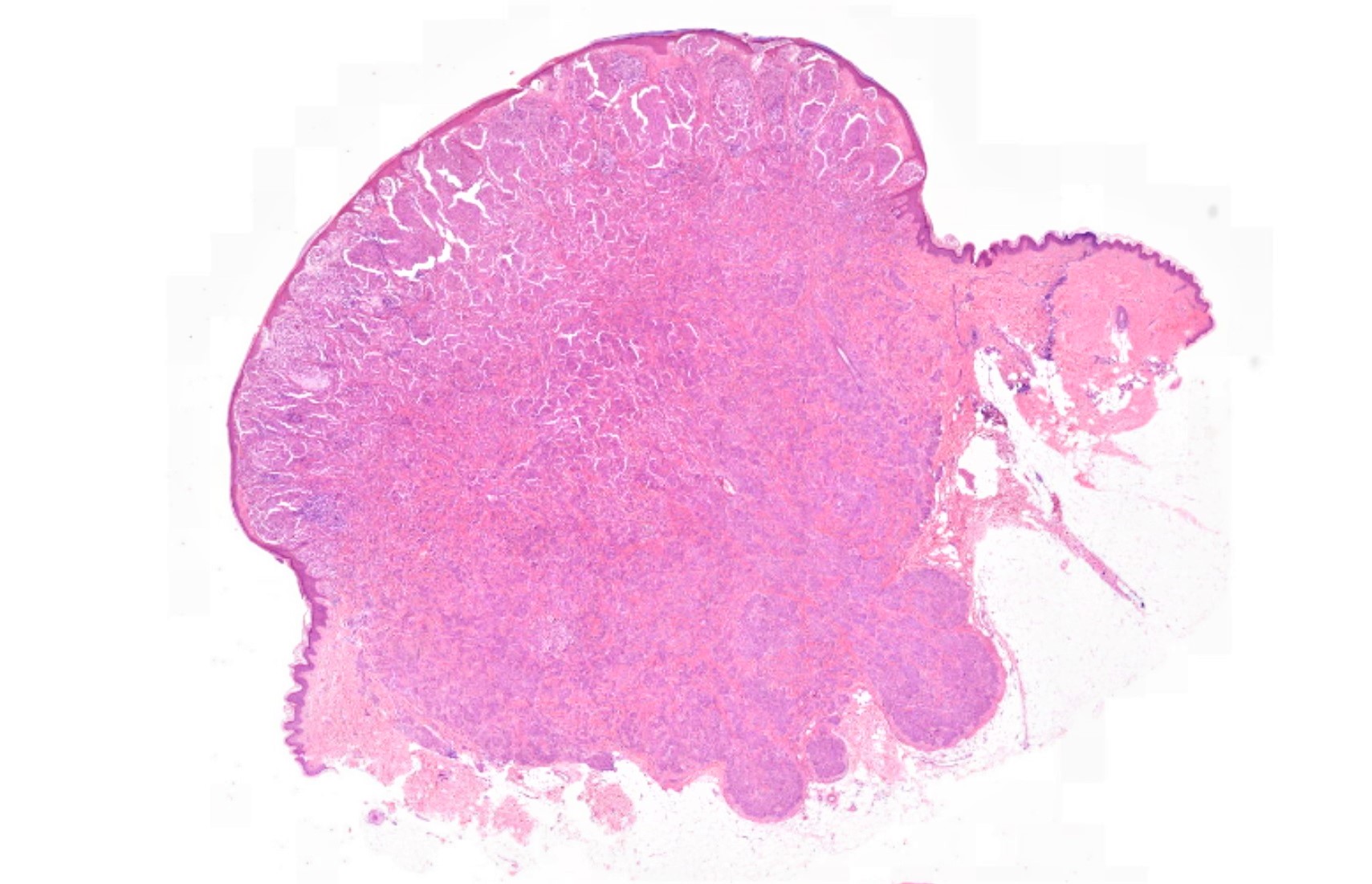
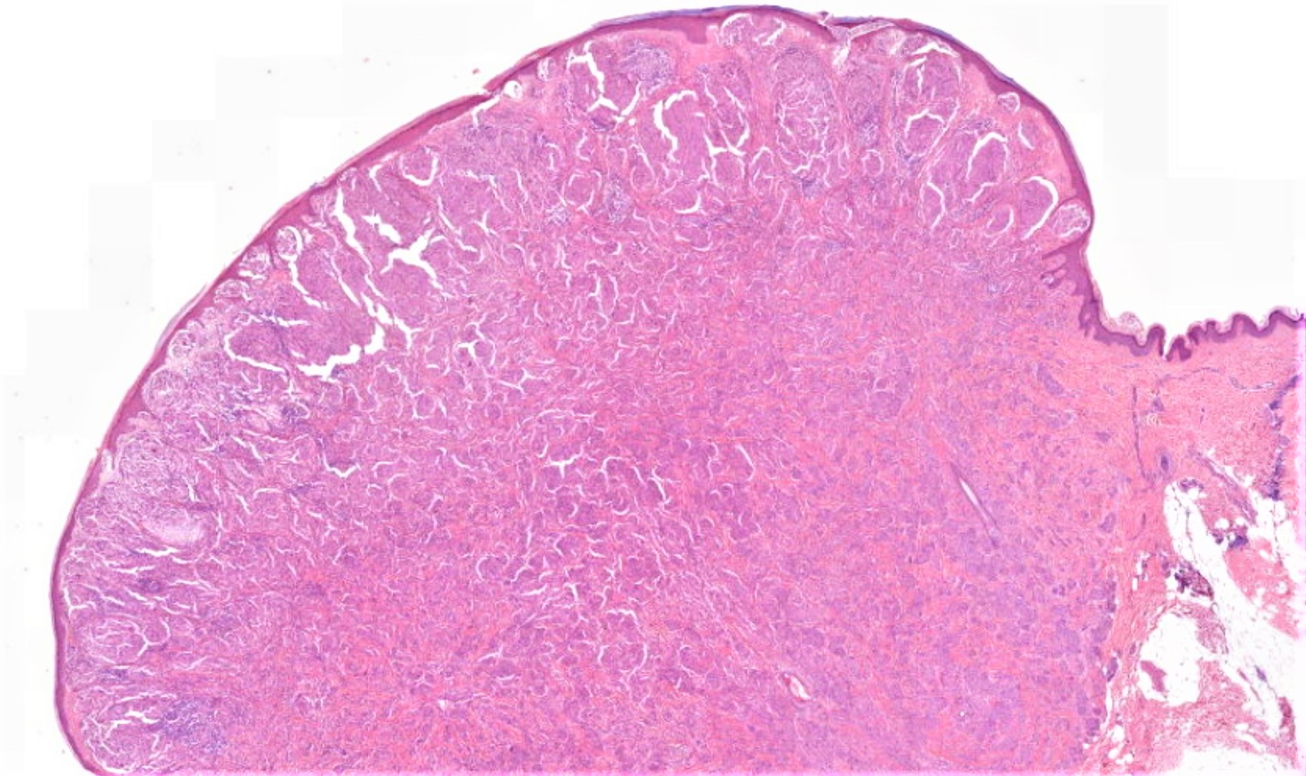
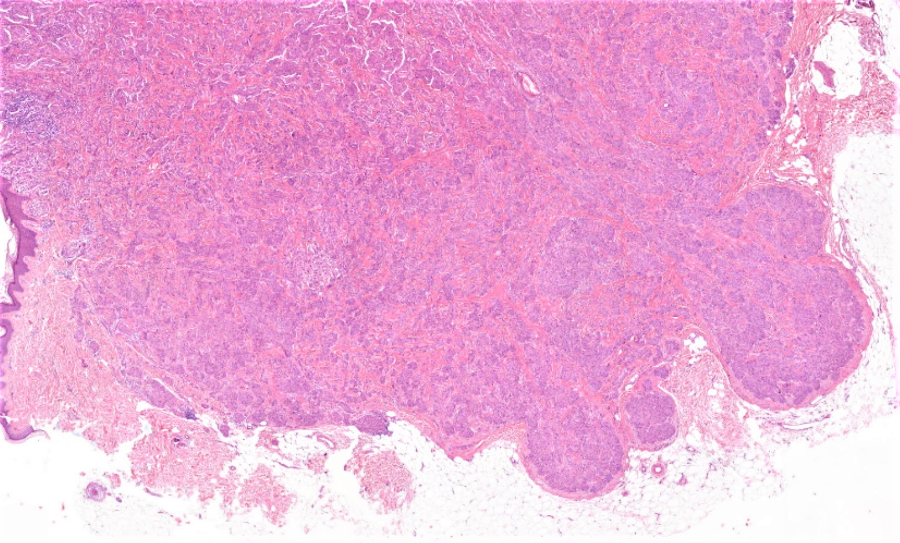
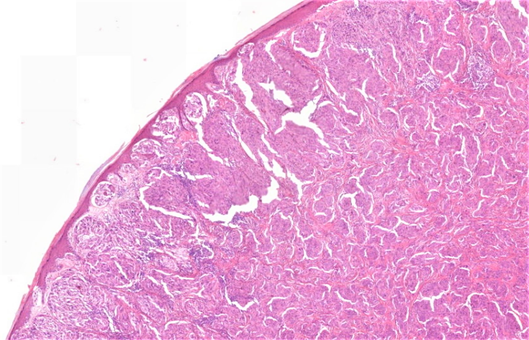
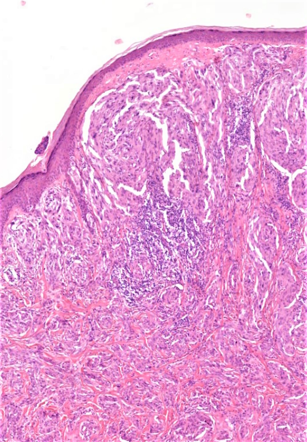
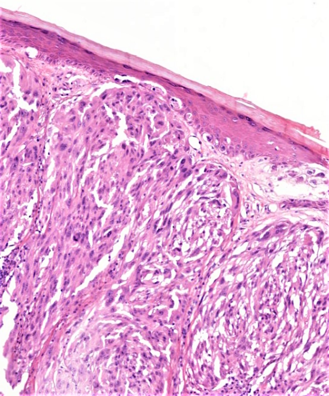
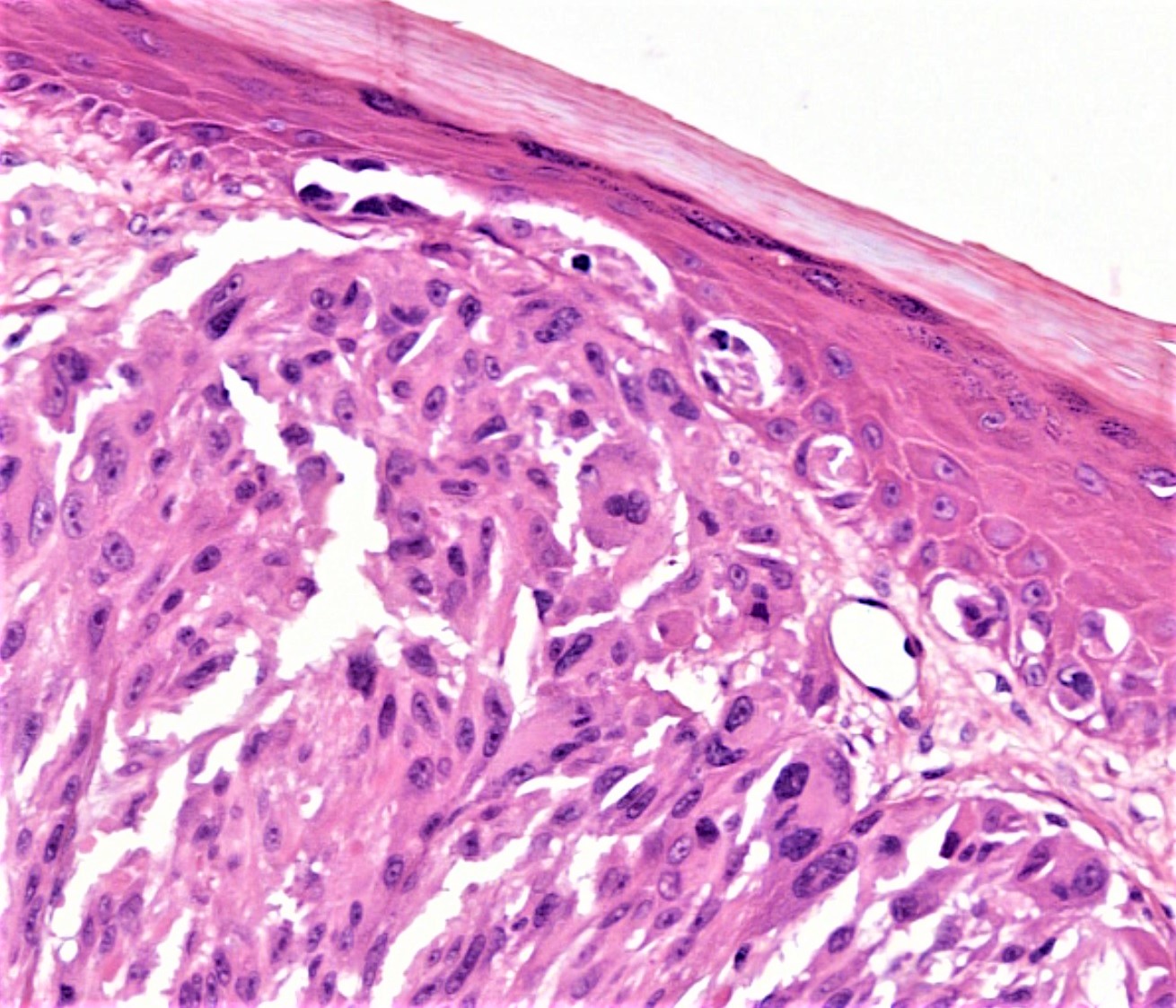
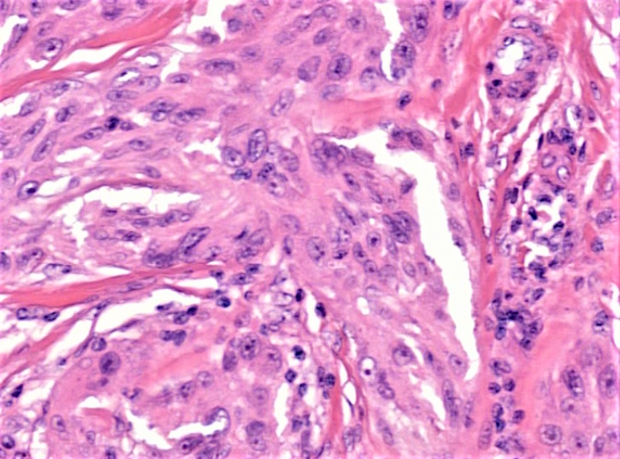
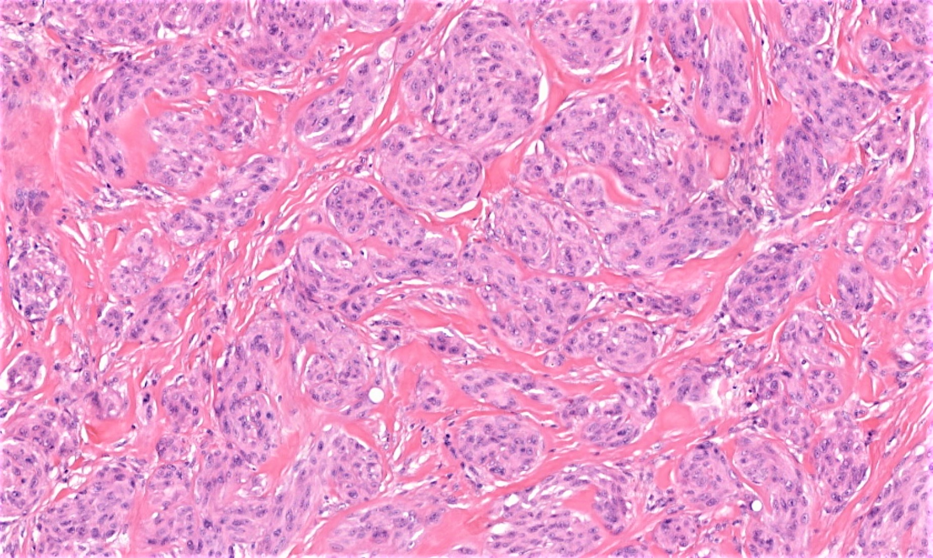
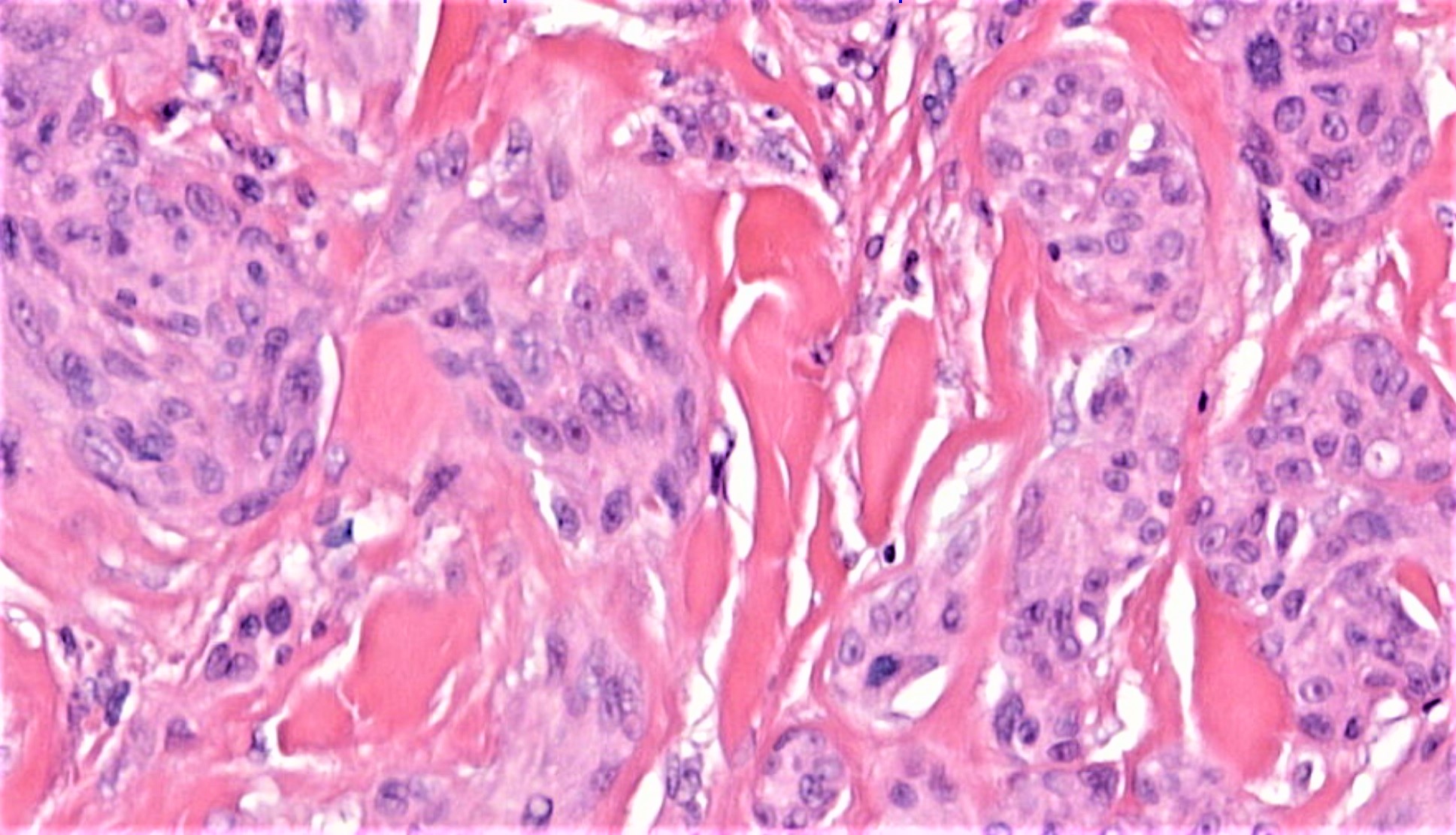
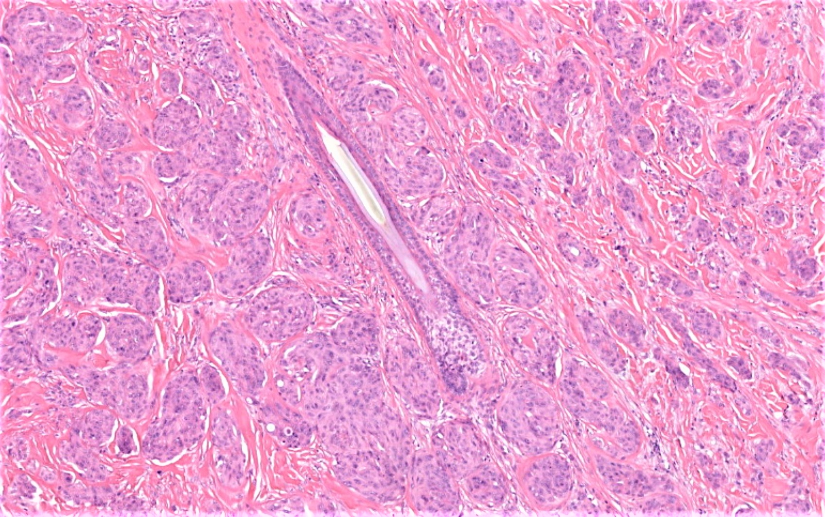
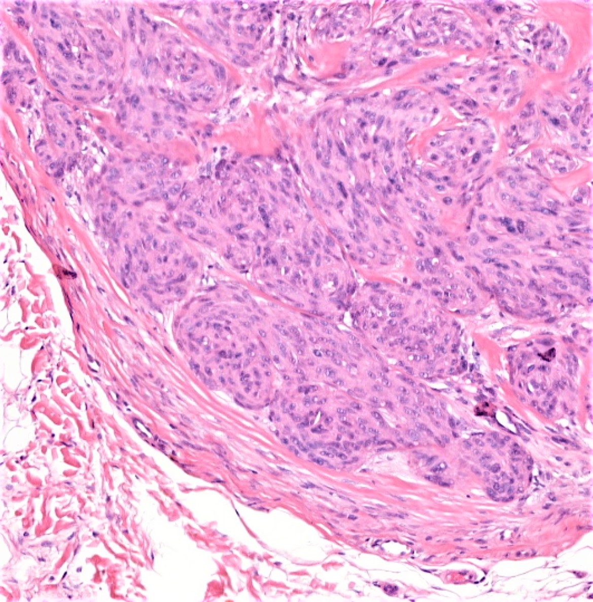
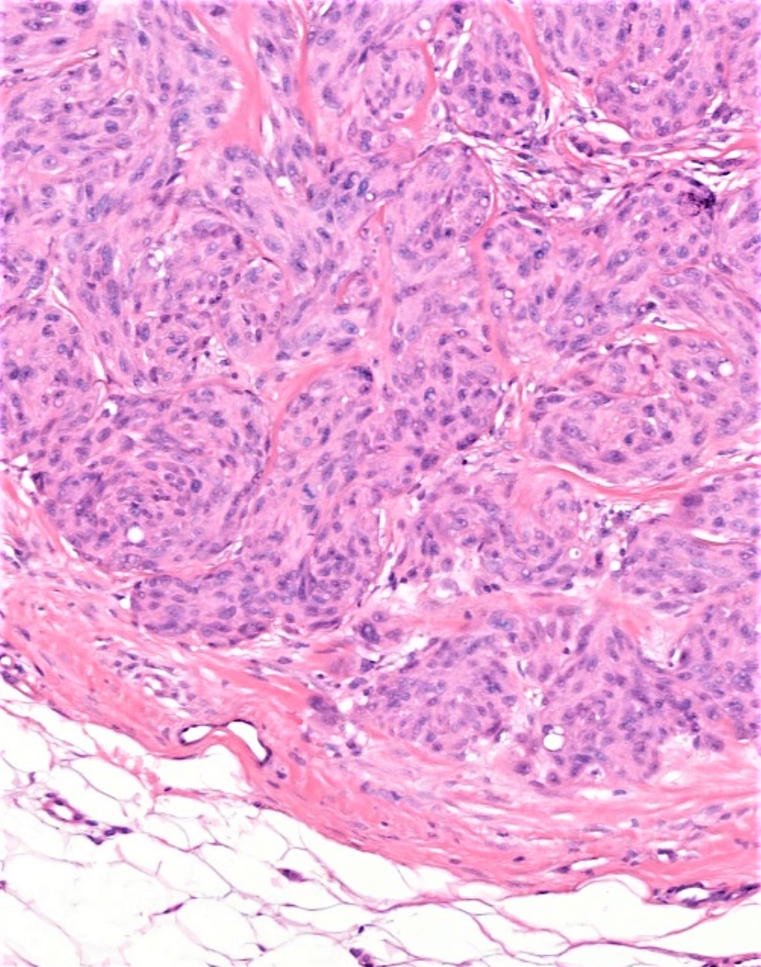
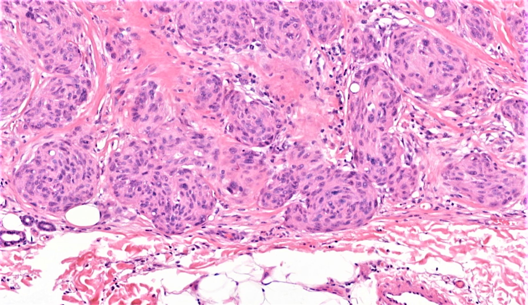
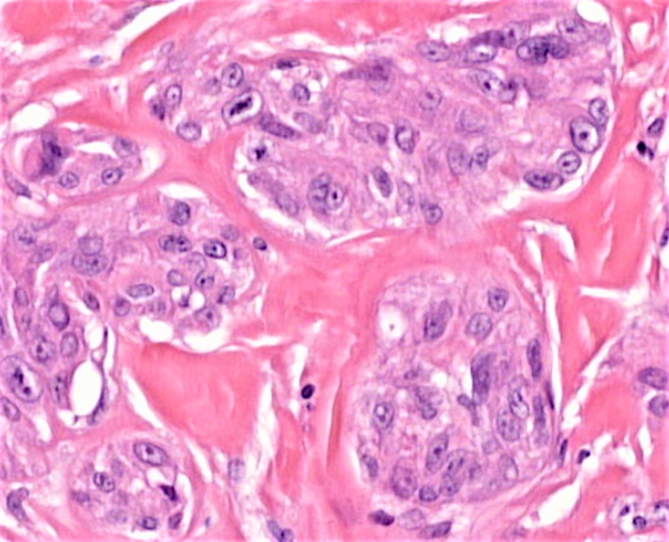
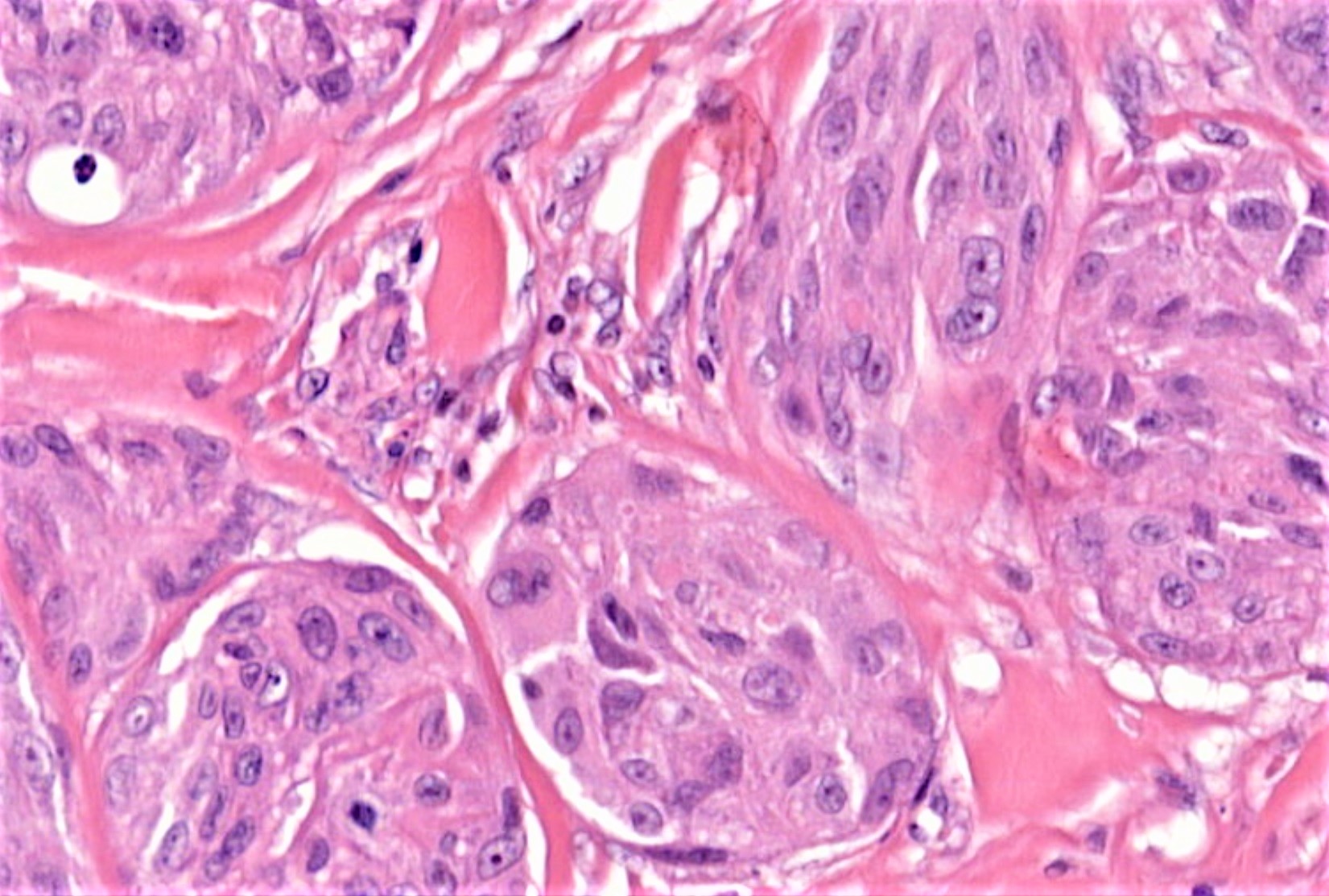
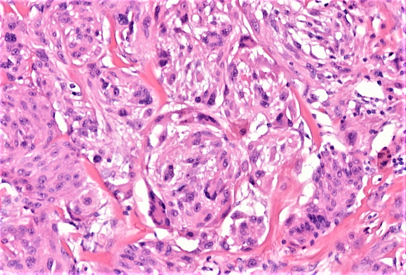
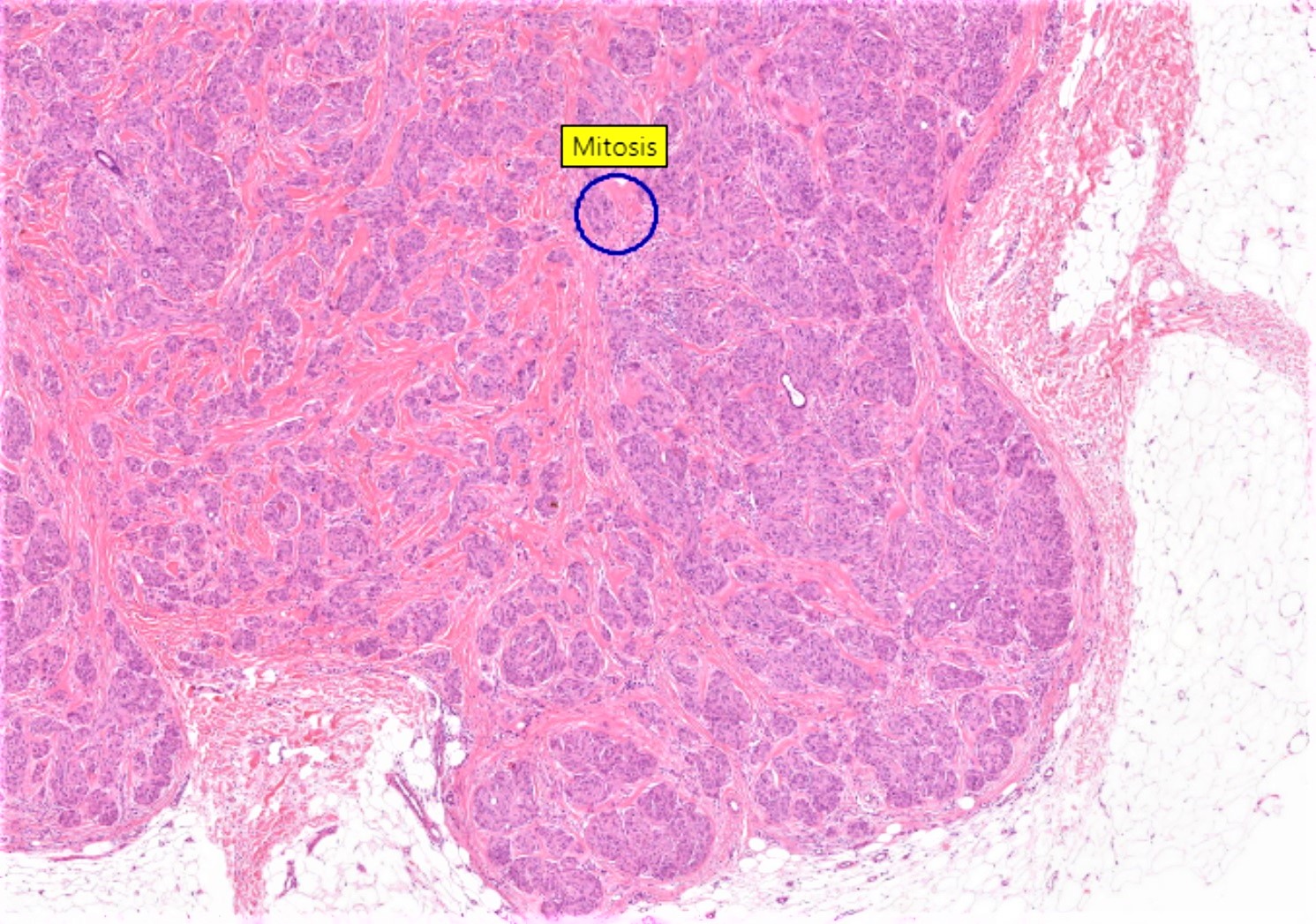
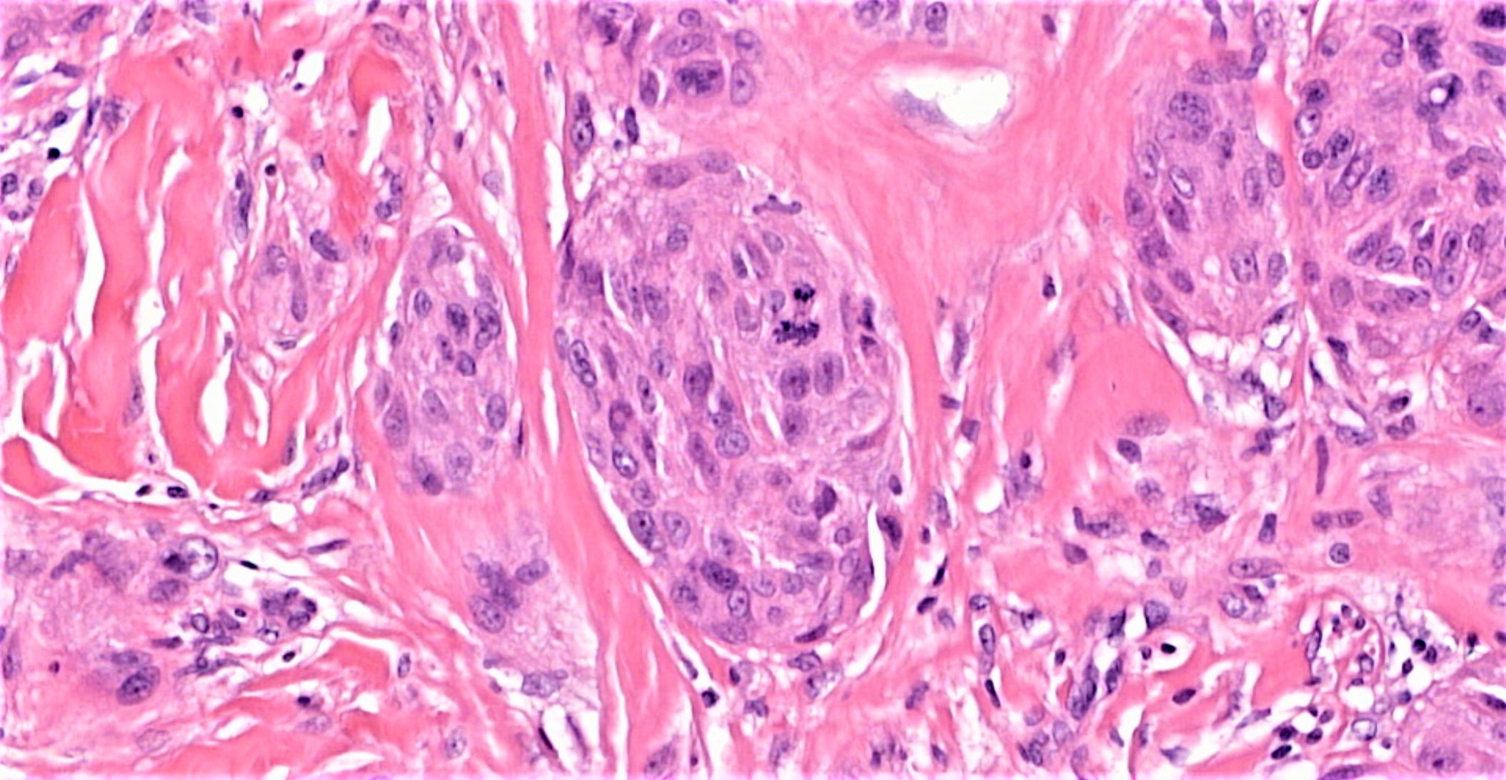
Diagnostic Pearls : Case 1869 - 27 July - Dr Arti Bakshi
Posted No value
Edited by Admin_Dermpath
1
Join the conversation
You can post now and register later. If you have an account, sign in now to post with your account.