Jump to content
In this section we have spot diagnoses posted on a daily basis since June 2010, now over 4000! You can review the archived cases and read the suggested diagnoses by users and the final comment by the contributors.
Case are uploaded each week day by 10 am UK time with the correct diagnosis will generally be posted at 8 pm UK time. Why not view the most recent spot diagnosis and proffer a diagnosis?

Case Number : Case 1202 - 30th January Posted By: Guest

Please read the clinical history and view the images by clicking on them before you proffer your diagnosis.
Submitted Date :
   (0 reviews)

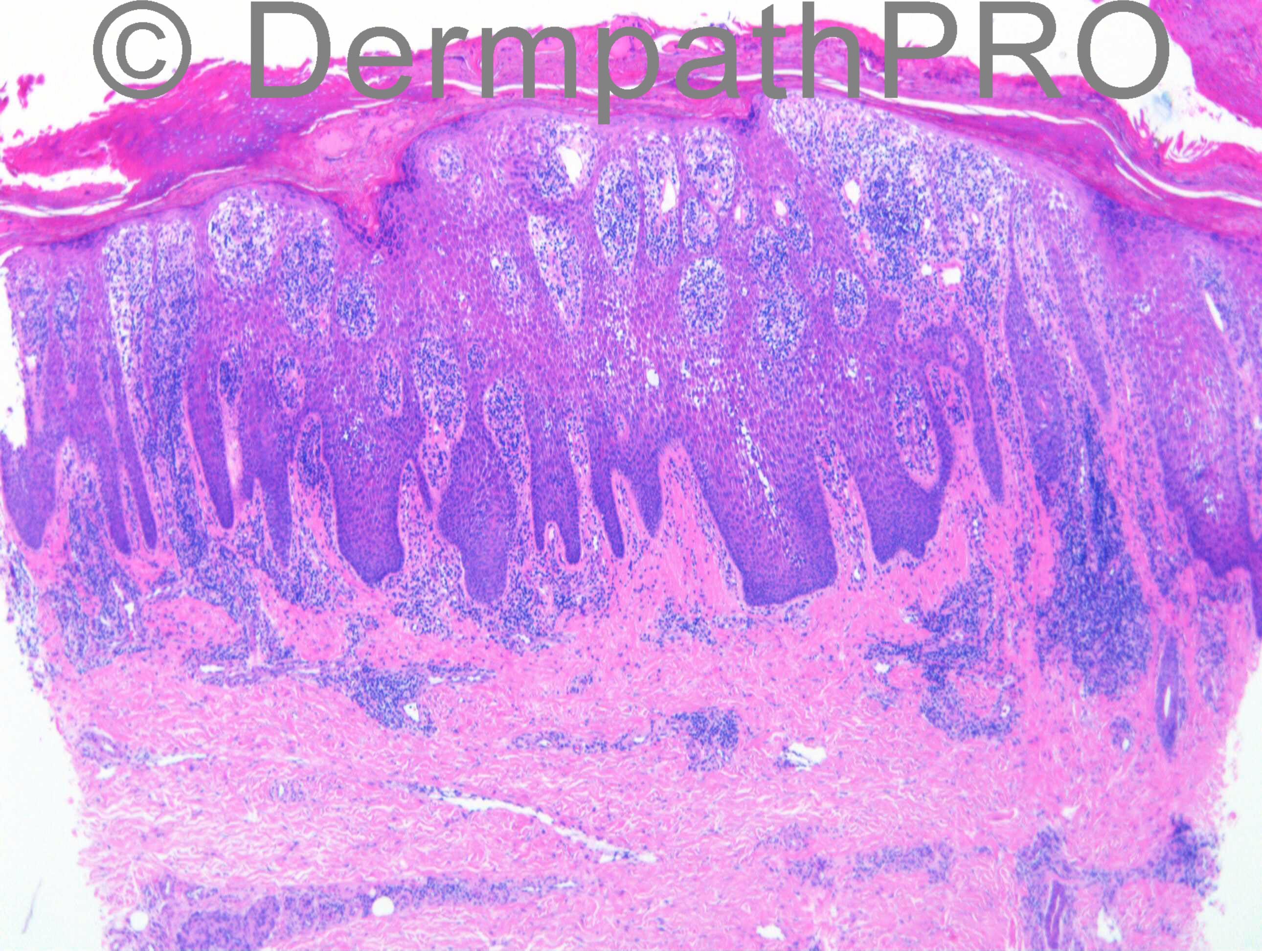
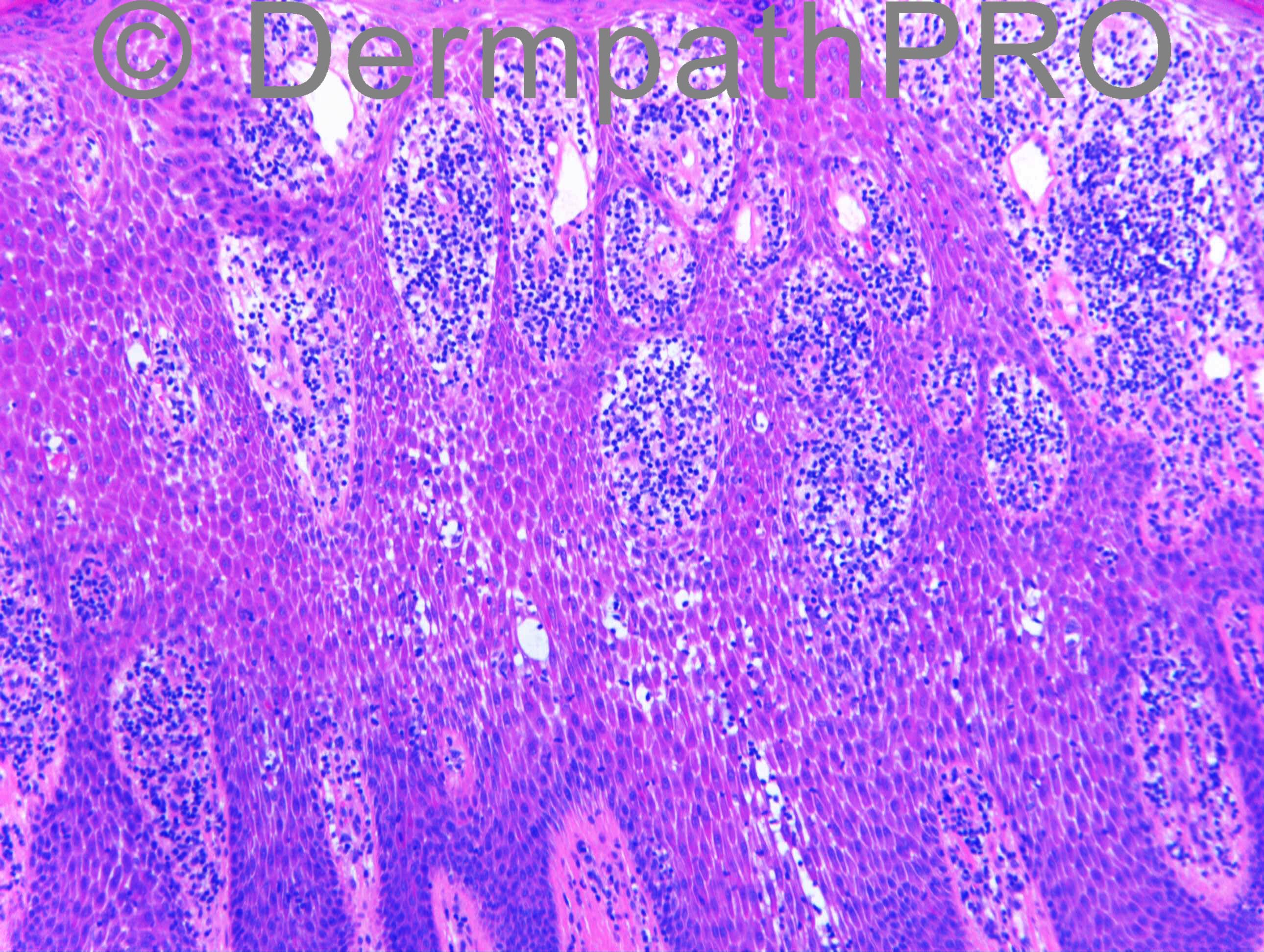
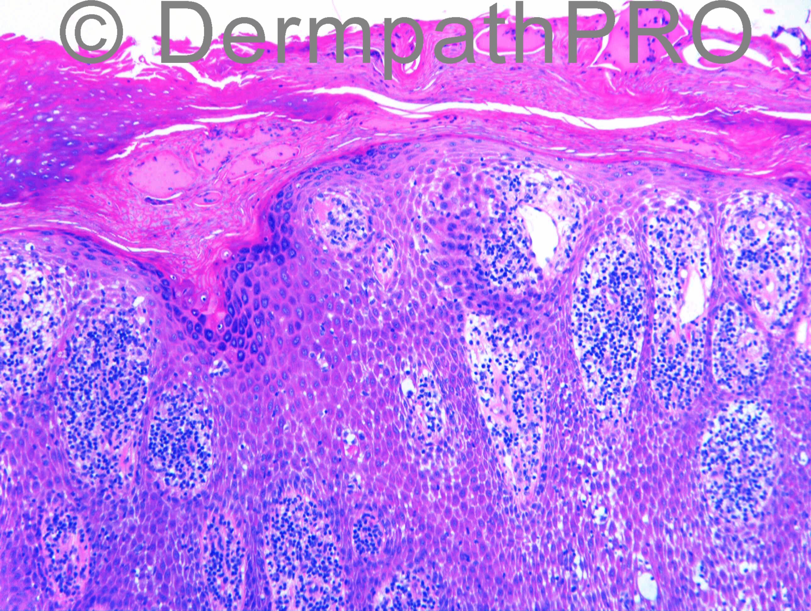
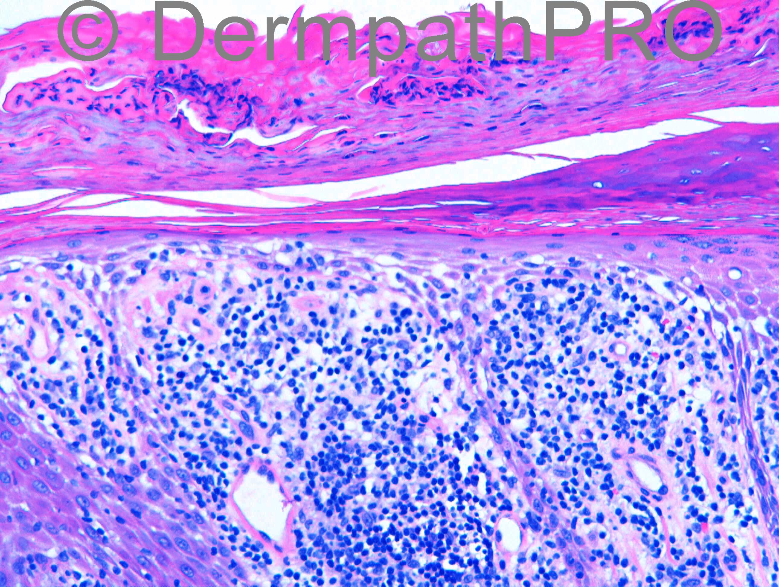
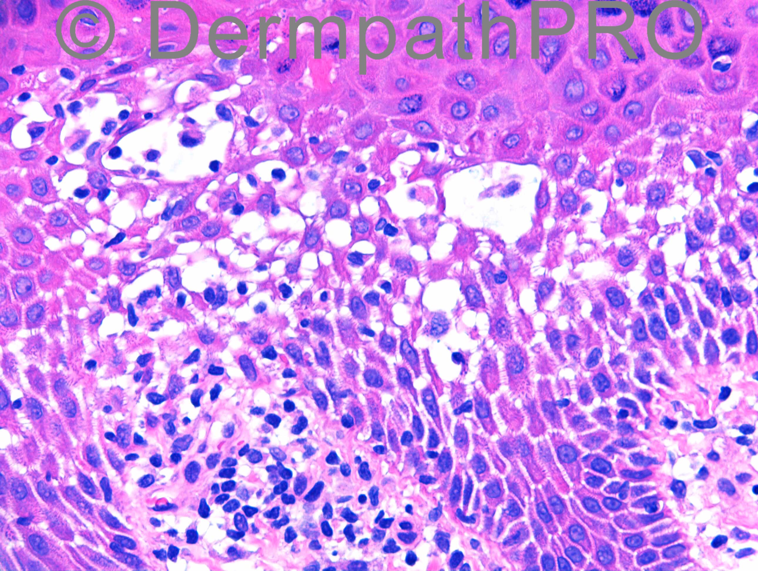
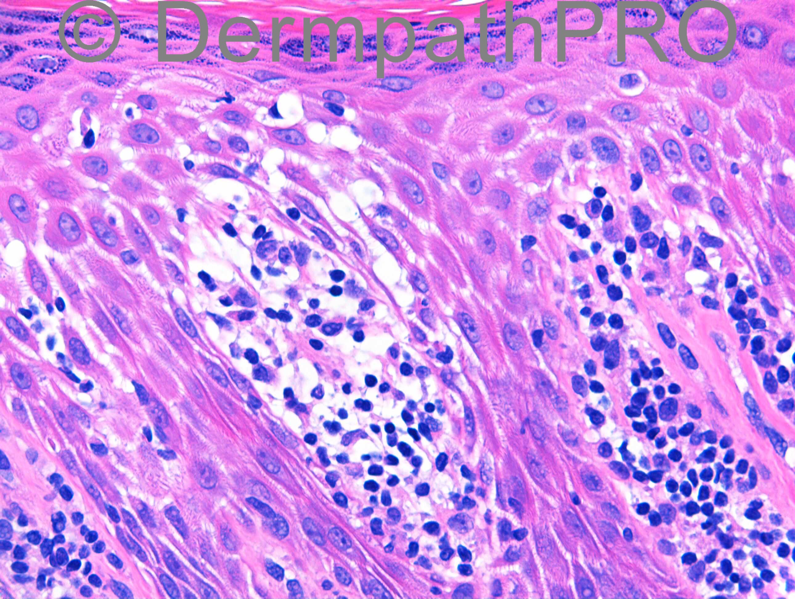
M31. Left palm biopsy. 3y well demarcated hyperkeratotic plaques on left palm and erythematous papules on left wrist and right shin. No nail psoriasis. DD: Psoriasis, eczema, CTCL

Case posted by Dr Richard Carr


  Report Record

User Feedback


Dr. Mona Abdel-Halim

Posted

I did not perceive the lymphocytes within the epidermis as malignant. I also see evident spongiosis mainly in the lower epidermis with stellate and elongated keratinocytes and vesicles. Parakeratosis with neutrophils. Areas of supra papillary thinning and some dilated capillaries. I think this is psoriasis, which on the palms and soles can be very difficult to differentiate from Eczema.

Share this comment


Link to comment
share_externally

Guest Romualdo

Posted

Agree with Mona. Almost all lymphocytes are located within dermal papillae. The ones within the epidermis are of the same size as dermal lymphocytes and do not look overtly malignant. Marked spongiosis militates against mycosis fungoides. I think this is psoriasis.

Share this comment


Link to comment
share_externally

Eman El-Nabarawy

Posted

Although many features are suggestive of psoriasis, hypergranulosis and atypical lymphocytes make me favor MF.

Share this comment


Link to comment
share_externally

Dr. Richard Carr

Posted

I think we need more commentators please

Share this comment


Link to comment
share_externally

Dr. Richard Carr

Posted

Thanks Mark. I did not work it up for MF but I had the advantage of a second biopsy from the wrist that also favoured eczema. Although I think even in the palmar biopsy I favoured eczematous dermatitis for the exact reasons highlighted perfectly by Mark. My report as follows:

A and B Eczematous reaction pattern with fairly dense superficial perivascular lymphocytic infiltrate. Eosinophils not notable. No lymphoid atypia. No fungi (PAS).

I was then asked a specific question from the clinical colleague as follows: "Could this be hyperkeratotic palmar eczema?"

My supplementary report as follows:
[size=3][color=#000000]Thank you for asking me to re-visit the specimens. There is a description in Weedon’s textbook (“hyperkeratotic dermatitis of the palms”) that matches perfectly with that seen in this case (prominent spongiosis, dense lymphocytic infiltrate with prominent exocytosis). There is quite a bit of parakeratosis in the palmar biopsy but the relatively preserved or thickened granular layer and degree of spongiosis led me to favour an eczematous dermatitis in this biopsy rather than psoriasis although the latter distinction can be exceedingly difficult in acral biopsies. The features in the biopsy from the wrist also favoured an eczematous dermatatitis rather than psoriasis.[/color][/size]

[size=4][color=#000000]Additional remarks: Agree about knee-jerk of doing PAS for fungi in eczematous reactions generally and especially localised lesions including particular anatomic locations (feet, hands, flexures and moist areas).[/color][/size]

Share this comment


Link to comment
share_externally


Join the conversation

You can post now and register later. If you have an account, sign in now to post with your account.

Guest
Add a comment...

×   Pasted as rich text.   Paste as plain text instead

  Only 75 emoji are allowed.

×   Your link has been automatically embedded.   Display as a link instead

×   Your previous content has been restored.   Clear editor

×   You cannot paste images directly. Upload or insert images from URL.

Loading...

×
×
  • Create New...