Jump to content
In this section we have spot diagnoses posted on a daily basis since June 2010, now over 4000! You can review the archived cases and read the suggested diagnoses by users and the final comment by the contributors.
Case are uploaded each week day by 10 am UK time with the correct diagnosis will generally be posted at 8 pm UK time. Why not view the most recent spot diagnosis and proffer a diagnosis?

Case Number : Case 1312 - 03 July Posted By: Guest

Please read the clinical history and view the images by clicking on them before you proffer your diagnosis.
Submitted Date :
   (0 reviews)

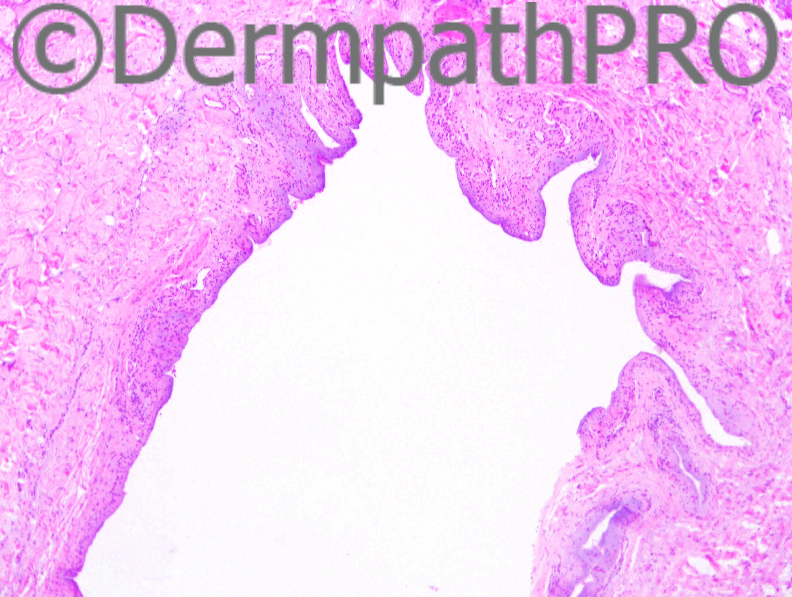
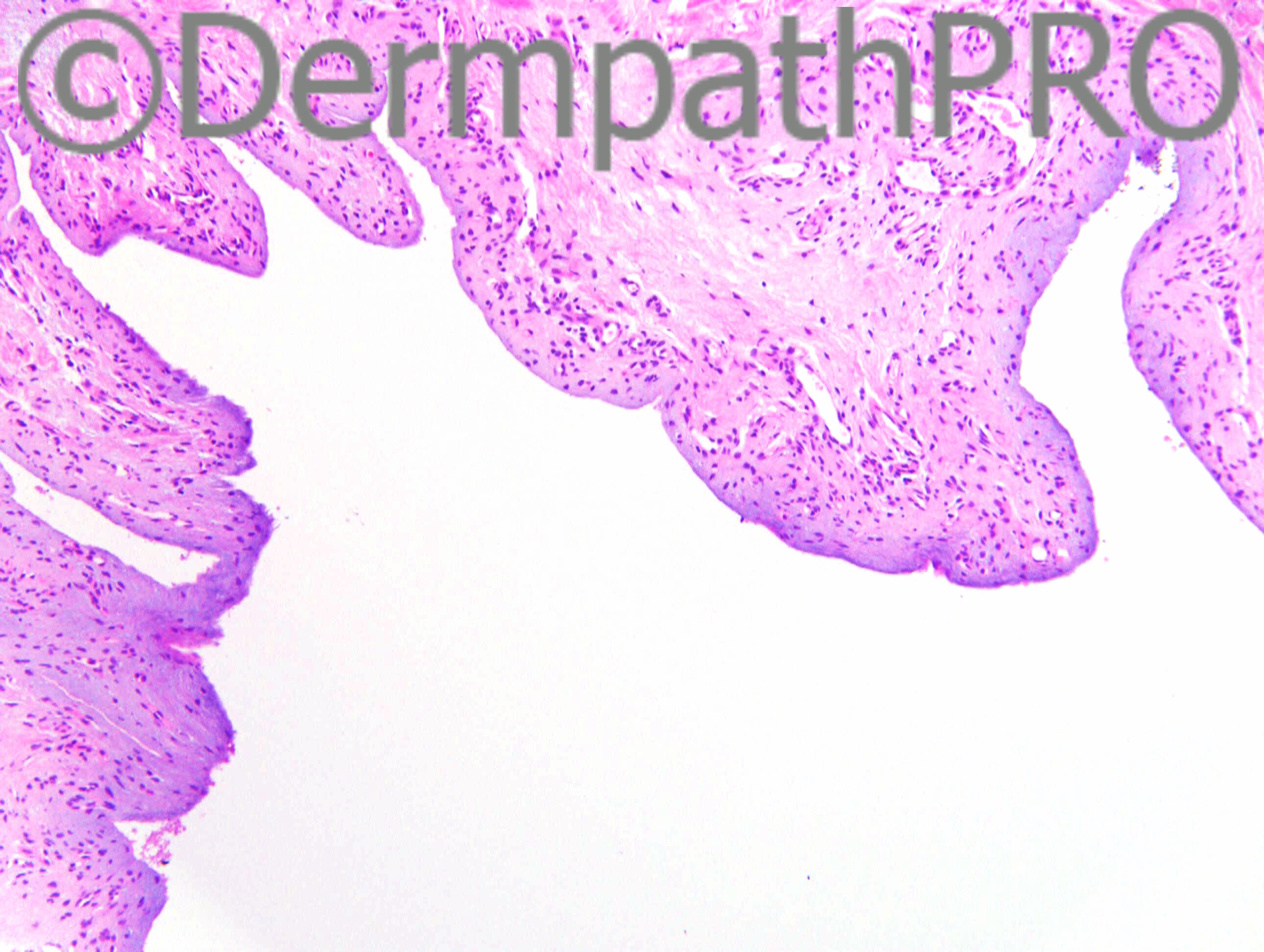
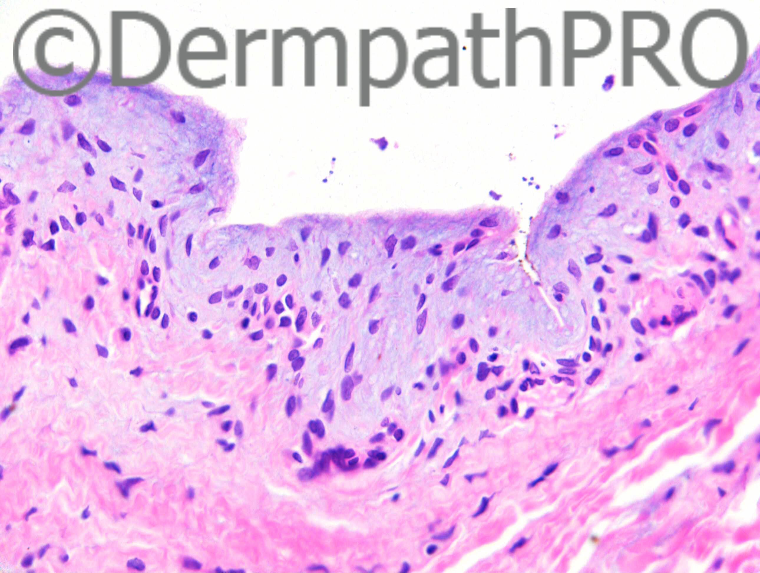
1cm soft cystic swelling on forehead ?sebaceous cyst

Case posted by Dr Richard Carr


  Report Record

User Feedback


Dr. Richard Carr

Posted

This is from a male of 53 years

Share this comment


Link to comment
share_externally

Dr. Mona Abdel-Halim

Posted

Metaplastic synovial cyst.

Share this comment


Link to comment
share_externally

Guest Romualdo

Posted

Yes: metaplastic synovial cyst.

Share this comment


Link to comment
share_externally

Eman El-Nabarawy

Posted

Cutaneous metaplastic synovial cyst.

Share this comment


Link to comment
share_externally

Dr. Richard Carr

Posted

[img]https://dermpathpro.com/uploads/spot_diagnosis_comment_img/Case%201312%20-%20RAC7159x10_4pm.jpg[/img]

[img]https://dermpathpro.com/uploads/spot_diagnosis_comment_img/Case%201312%20-%20RAC7159x20_4pm.jpg[/img]

[img]https://dermpathpro.com/uploads/spot_diagnosis_comment_img/Case%201312%20-%20RAC7159x40_4pm.jpg[/img]

[img]https://dermpathpro.com/uploads/spot_diagnosis_comment_img/Case%201312%20-%20RAC7159x5b_4pm.jpg[/img]

Share this comment


Link to comment
share_externally

Dr. Mona Abdel-Halim

Posted

OMG :-))
So it is just a cystic BCC that fell out ..
I thought of this when Dr Carr said try again !!!! Was going to suggest it but the outline of the cavity (festooning outline) was holding me back !!!
Looking again to the initial images I think the mucin in the last image should have been taken as a clue ...
Lovely :-))

Share this comment


Link to comment
share_externally

Dr. Richard Carr

Posted

[img]https://dermpathpro.com/uploads/spot_diagnosis_comment_img/Case%201312%20-%20RAC7159x5_BerEP4_5pm.jpg[/img]

Share this comment


Link to comment
share_externally

Dr. Mona Abdel-Halim

Posted

Just amazing :-))

Share this comment


Link to comment
share_externally

Dr. Richard Carr

Posted

Yes I agree it is a terrible trick but I can assure you several years ago I reviewed a biopsy from the foot (not a common site for BCC) in which the report read roughly as follows: "Dermal cyst and a separate carry-over of nodular BCC". On review the cyst had a mucinous stroma but no epithelial lining and the so-called carry-over was a similar shape to the hole in the dermis! I have recorded two further examples in my database in which I reported punch biopsies from the face clinically ?BCC with just a mucinous pseudocyst such as this and could not find the BCC despite levels through the block - I reported the biopsies as most likely a BCC that has "dropped-out". It is a trick well to remember!

Share this comment


Link to comment
share_externally


Join the conversation

You can post now and register later. If you have an account, sign in now to post with your account.

Guest
Add a comment...

×   Pasted as rich text.   Paste as plain text instead

  Only 75 emoji are allowed.

×   Your link has been automatically embedded.   Display as a link instead

×   Your previous content has been restored.   Clear editor

×   You cannot paste images directly. Upload or insert images from URL.

Loading...

×
×
  • Create New...