Jump to content
In this section we have spot diagnoses posted on a daily basis since June 2010, now over 4000! You can review the archived cases and read the suggested diagnoses by users and the final comment by the contributors.
Case are uploaded each week day by 10 am UK time with the correct diagnosis will generally be posted at 8 pm UK time. Why not view the most recent spot diagnosis and proffer a diagnosis?

Case Number : Case 1282 - 22 May Posted By: Guest

Please read the clinical history and view the images by clicking on them before you proffer your diagnosis.
Submitted Date :
   (0 reviews)

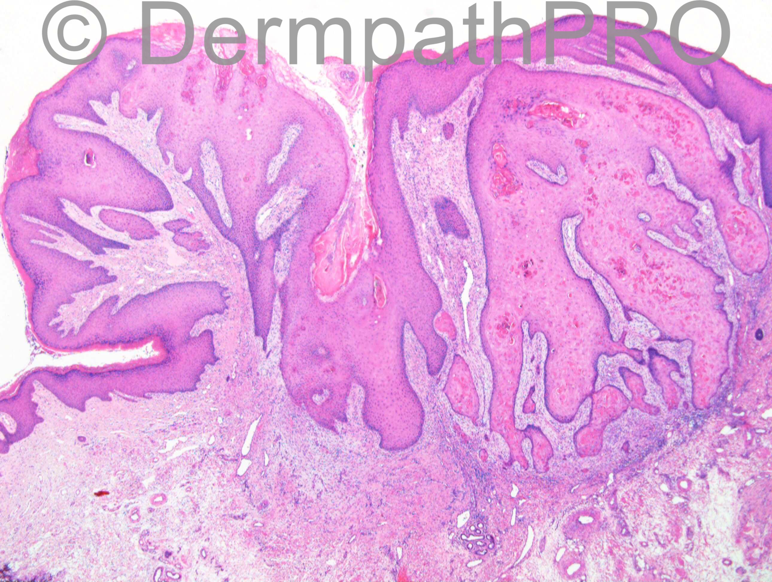
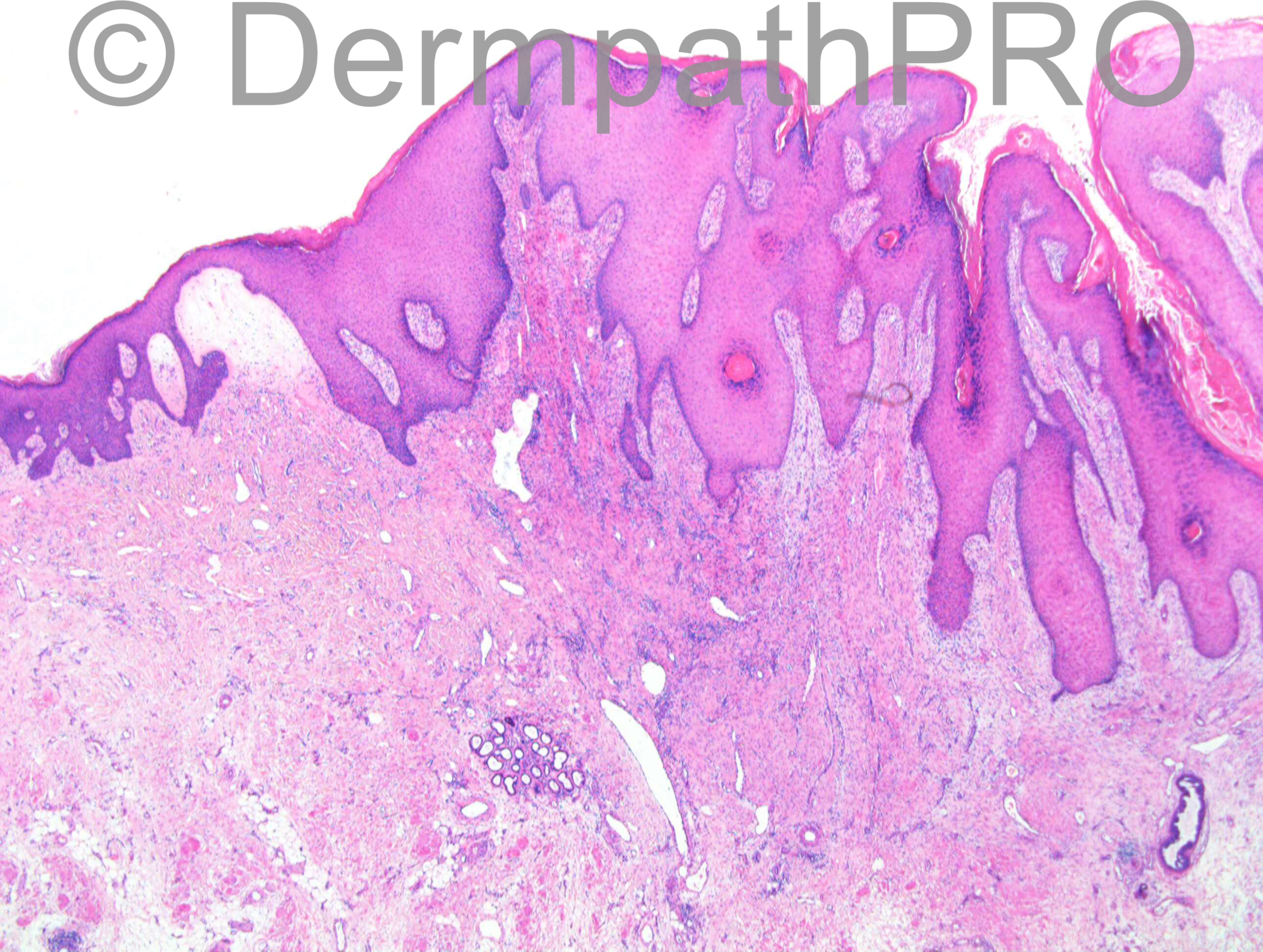
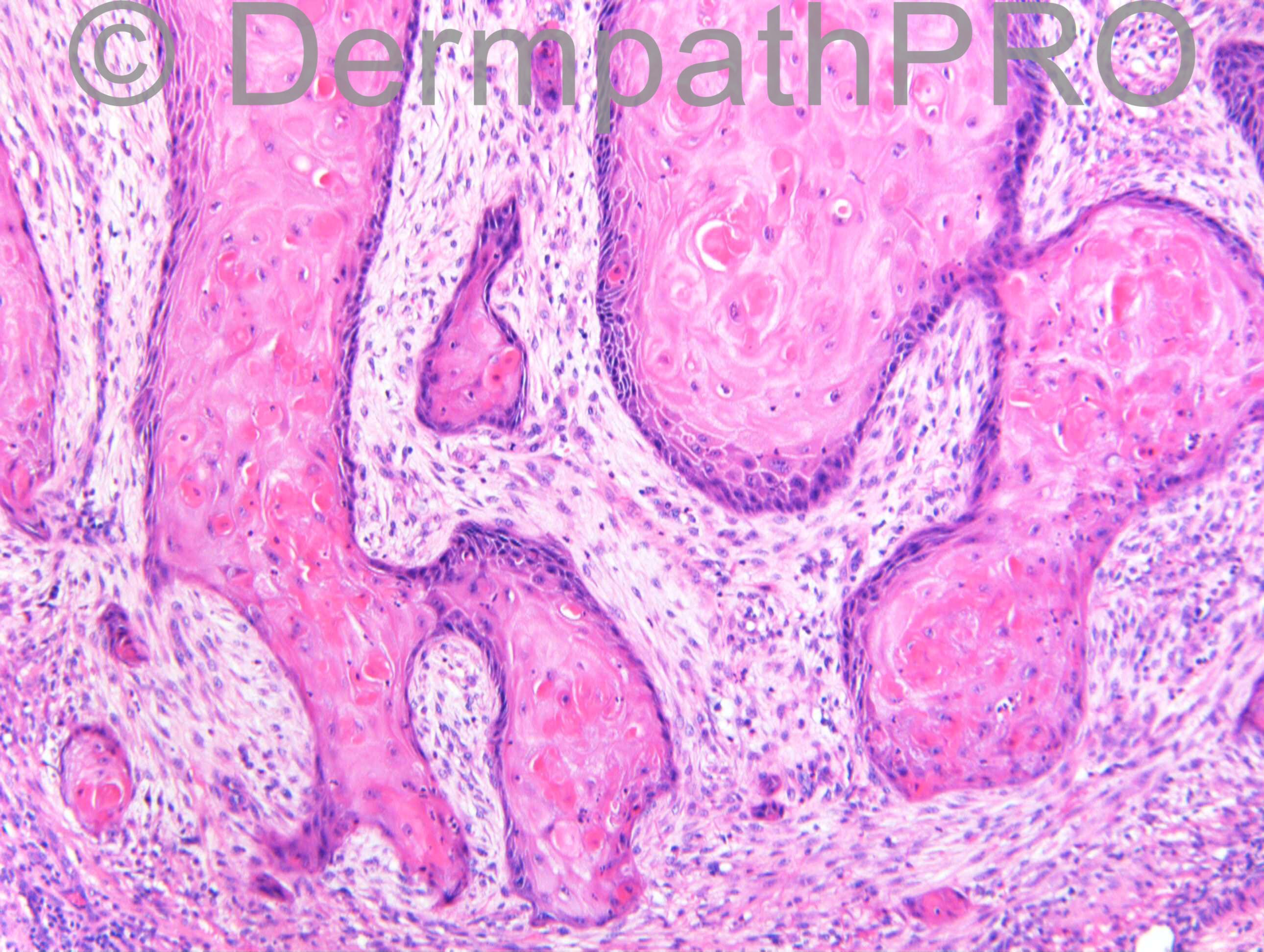
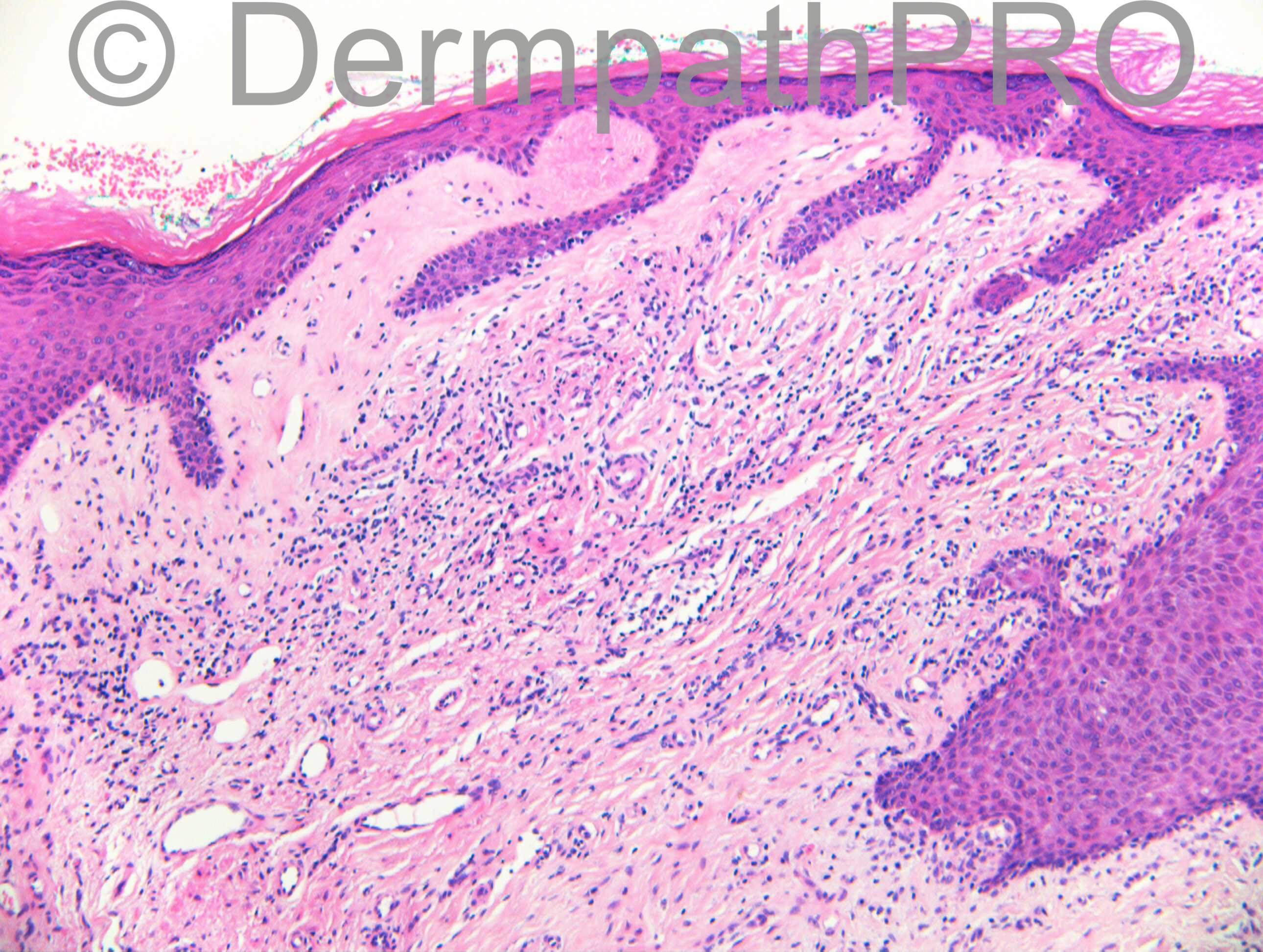
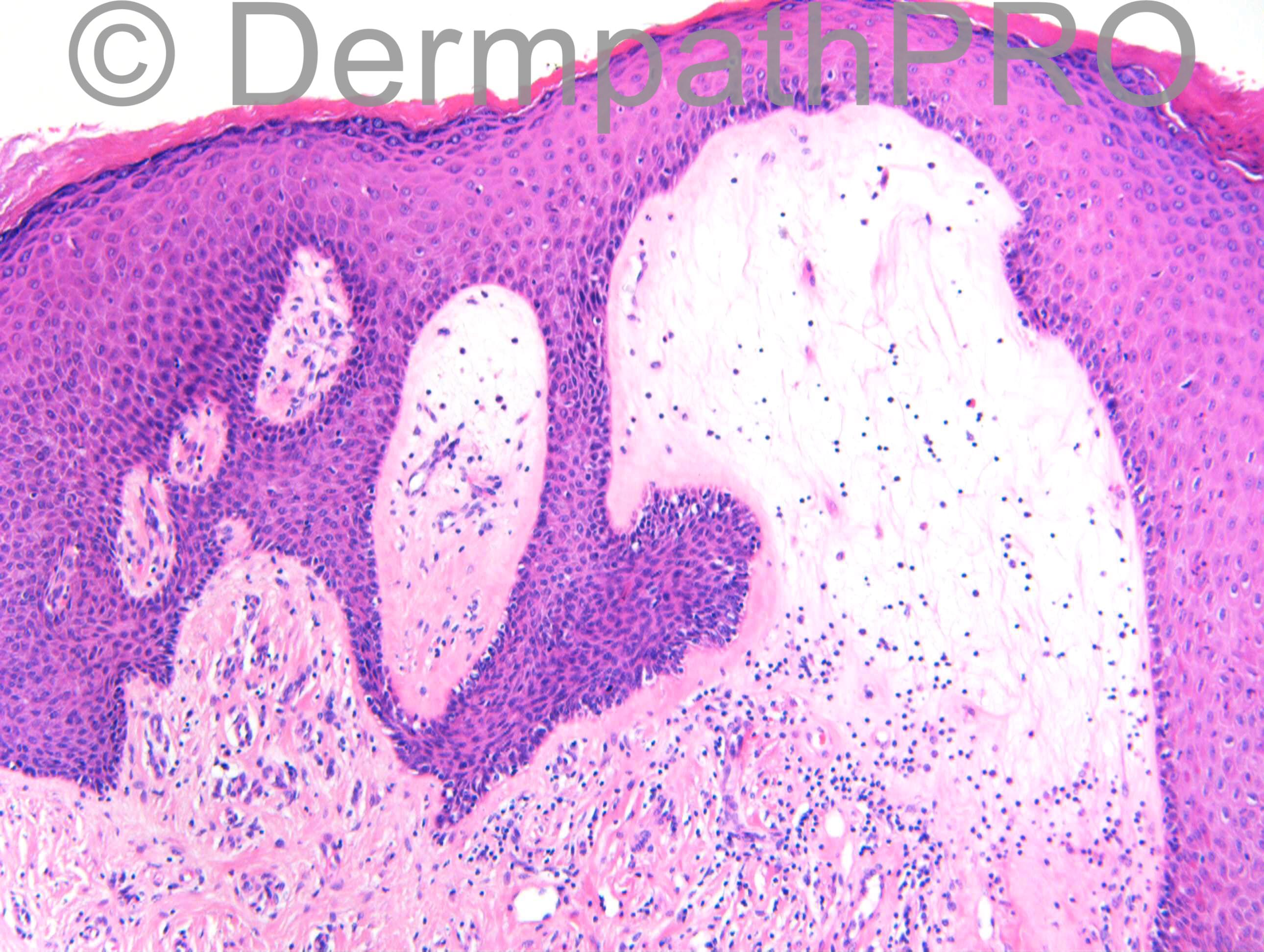
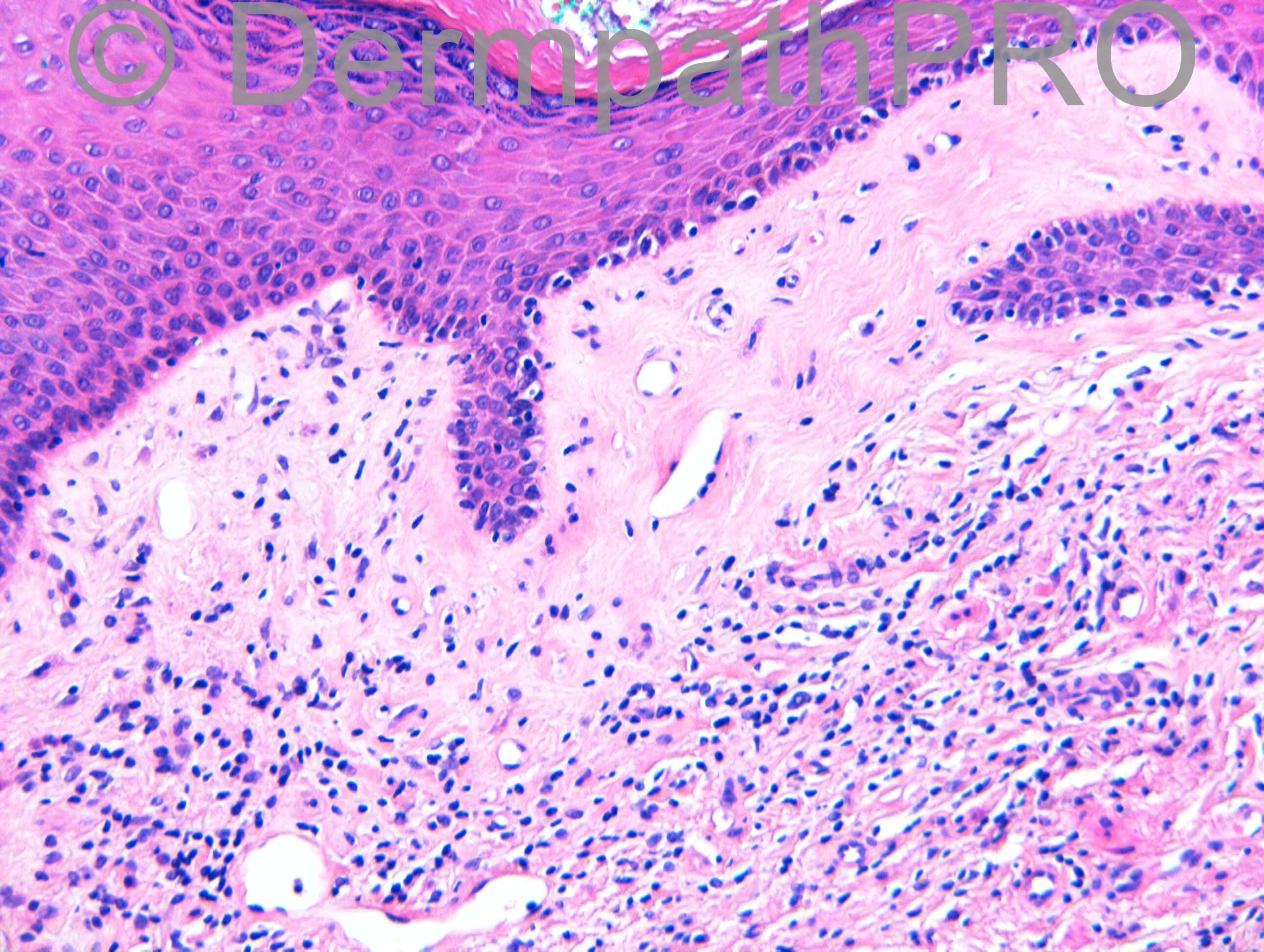
F89. No accurate history. ?3/12 6mm2 lesion at anal margin. ?SCC

Case posted by Dr Richard Carr

  Report Record

User Feedback


Guest Romualdo

Posted

Squamous cell carcinoma associated with lichen sclerosus.

Share this comment


Link to comment
share_externally

IgorSC

Posted

Agree. Although the literature describes this association I really don´t see it consistently. I have only one case of SCC associated with lichen sclerosus among numerous cases of LE. What are yours experiences?

Share this comment


Link to comment
share_externally

Dr. Mona Abdel-Halim

Posted

I will call this follicular SCC arising from a precursor differentiated intraepithelial neoplasia complicating LS.

Share this comment


Link to comment
share_externally

Guest Tiberiu Tebeica

Posted

My thought was the same: SCC on LS

Share this comment


Link to comment
share_externally

Dr. Richard Carr

Posted

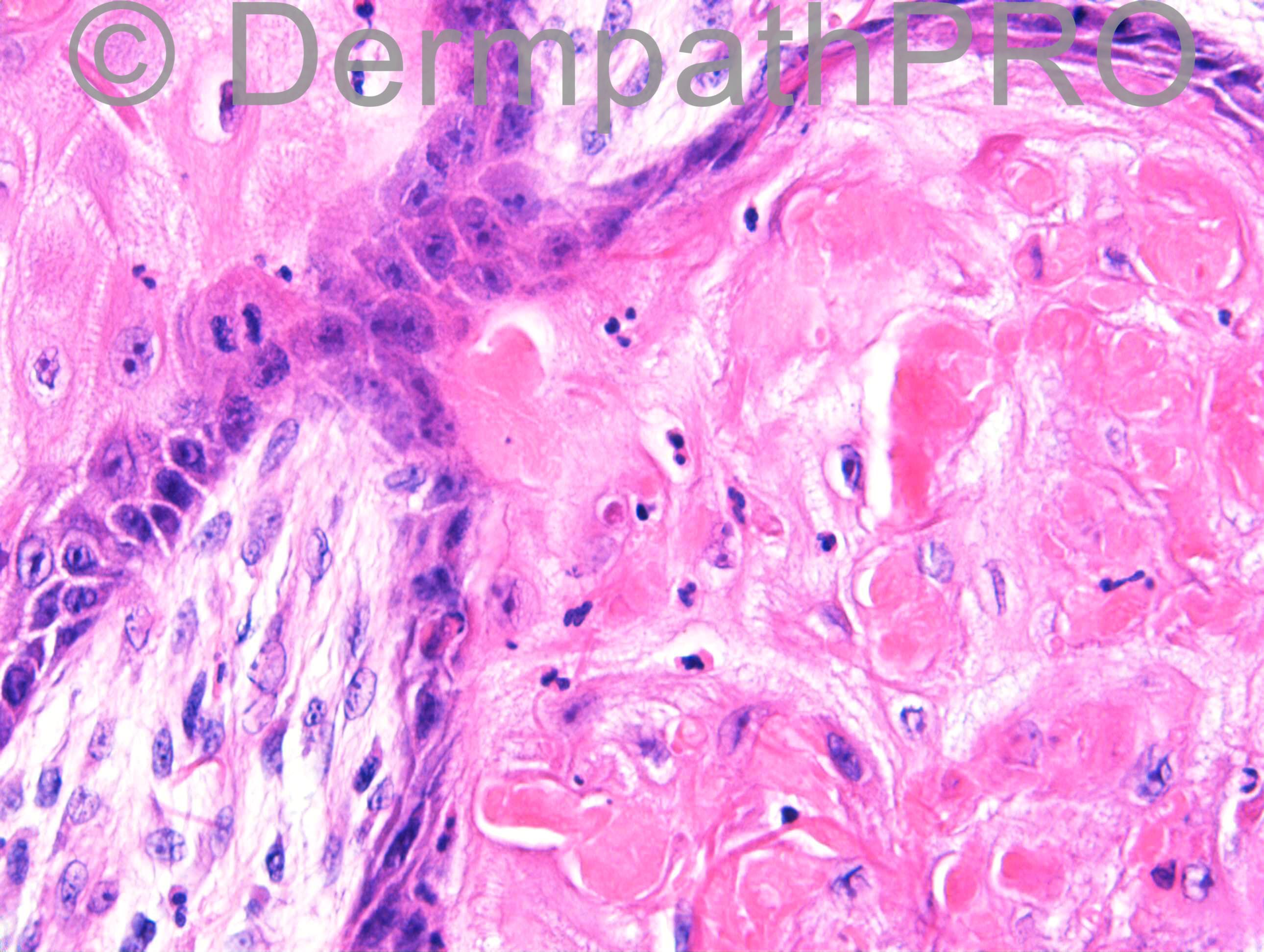
I reported this as [b][u]in keeping with [/u][/b]a keratoacanthoma and noted the background lichen sclerosus. Please note the elastic entrapment in last image within mature epithelium highly typical of KA. There is also scarring with vascular proliferation below the lesion typical of a late largely regressed lesion. The history was not perfect but seemed to fit in any case. I would only expect to see elastic entrapment within the mature epithelium in reactive follicular pseudoepitheliomatous conditions or KA but certainly not in my concept of a well differentiated squamous cell carcinoma (that would also not have a very short history).

Share this comment


Link to comment
share_externally

Dr. Richard Carr

Posted

I feel the need to change my diagnosis on this case in favour of a well differentiated squamous cell carcinoma, ano-genital type, resembling KA (or with KA-like features if you prefer). I think it can be exceedingly difficult (see Spot Diagnosis 1805, April 2017) but I think experience has affected my view of this older case and I should have re-commented earlier!  I have and will be very wary about making a diagnosis of KA on anogenital skin in future.

Share this comment


Link to comment
share_externally


Join the conversation

You can post now and register later. If you have an account, sign in now to post with your account.

Guest
Add a comment...

×   Pasted as rich text.   Paste as plain text instead

  Only 75 emoji are allowed.

×   Your link has been automatically embedded.   Display as a link instead

×   Your previous content has been restored.   Clear editor

×   You cannot paste images directly. Upload or insert images from URL.

Loading...

×
×
  • Create New...