Jump to content
In this section we have spot diagnoses posted on a daily basis since June 2010, now over 4000! You can review the archived cases and read the suggested diagnoses by users and the final comment by the contributors.
Case are uploaded each week day by 10 am UK time with the correct diagnosis will generally be posted at 8 pm UK time. Why not view the most recent spot diagnosis and proffer a diagnosis?

Case Number : Case 1466 -06 February Posted By: Guest

Please read the clinical history and view the images by clicking on them before you proffer your diagnosis.
Submitted Date :
   (0 reviews)

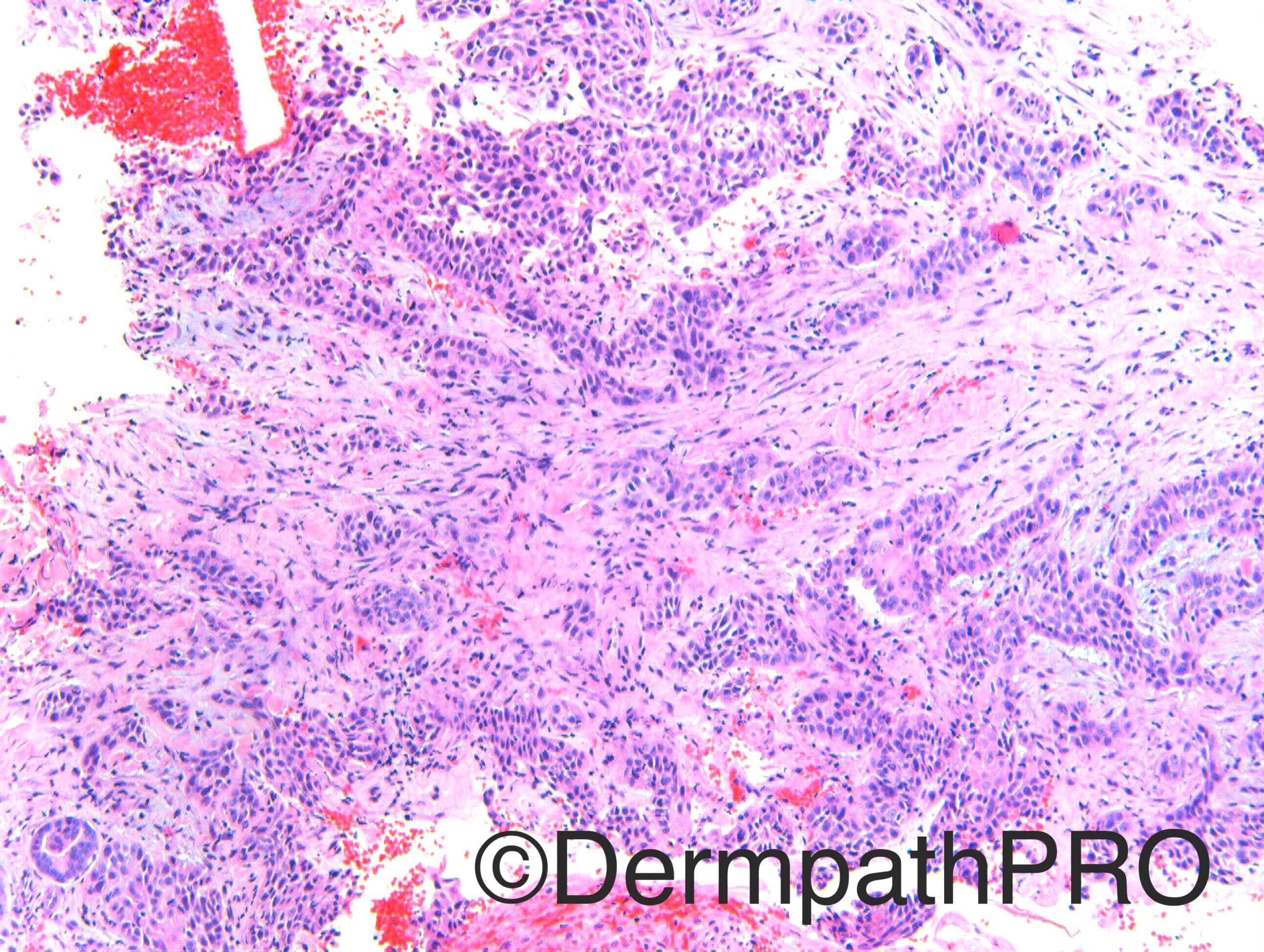
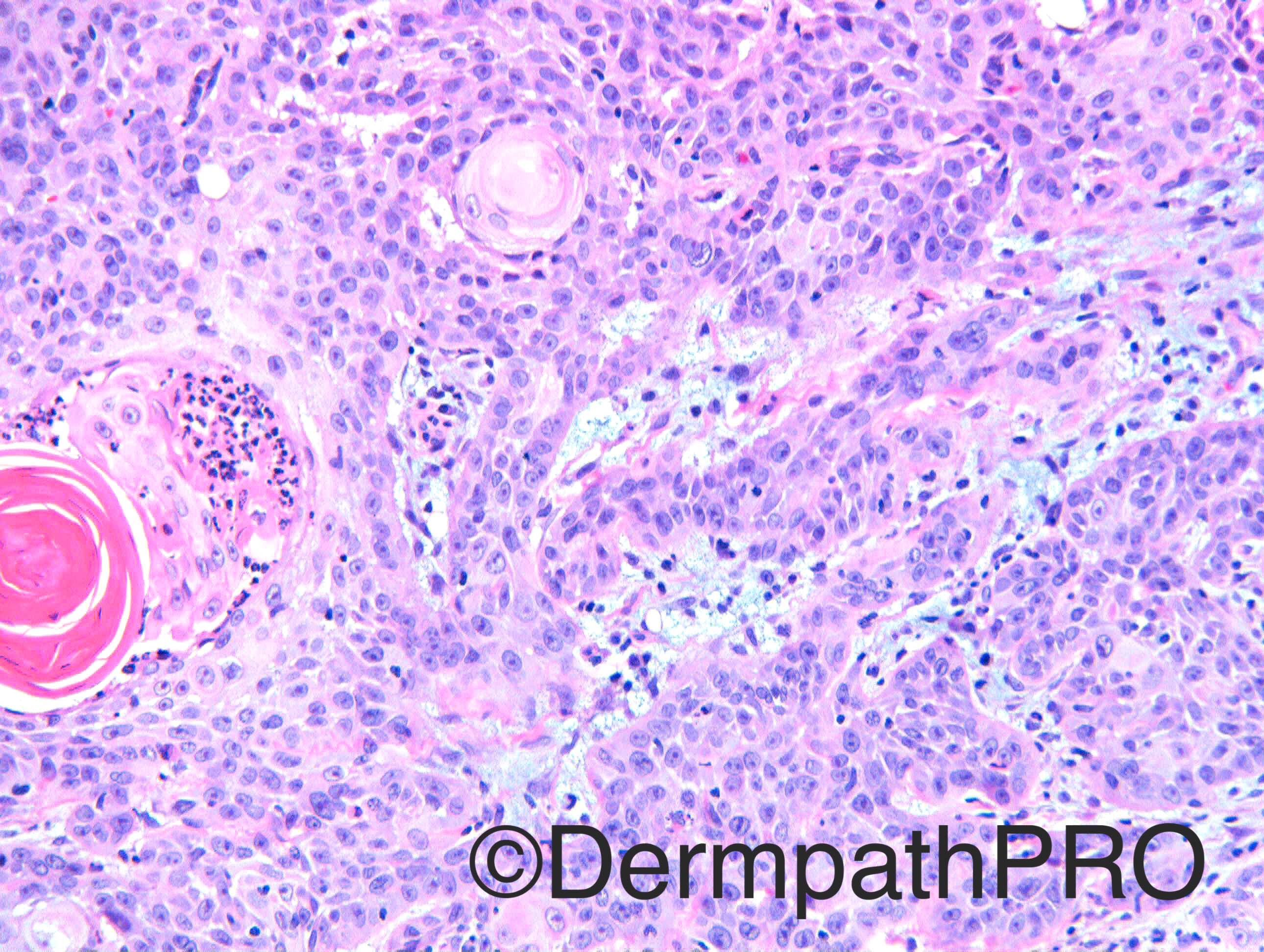
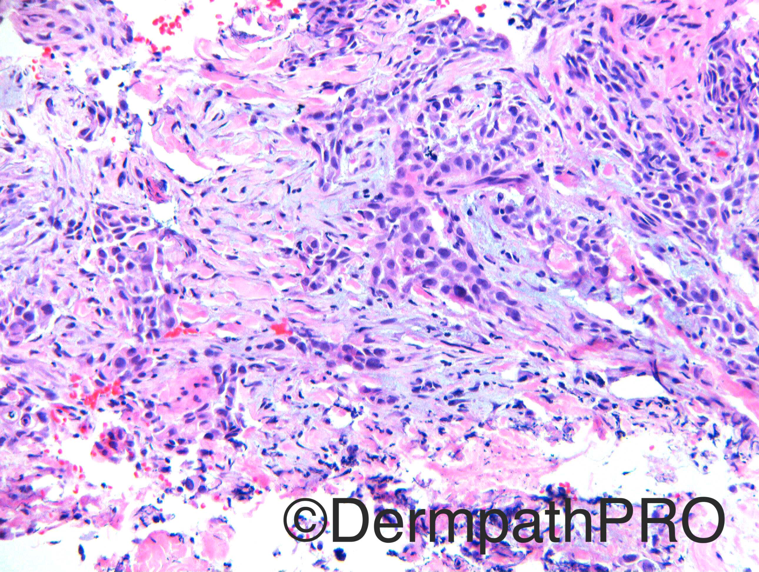
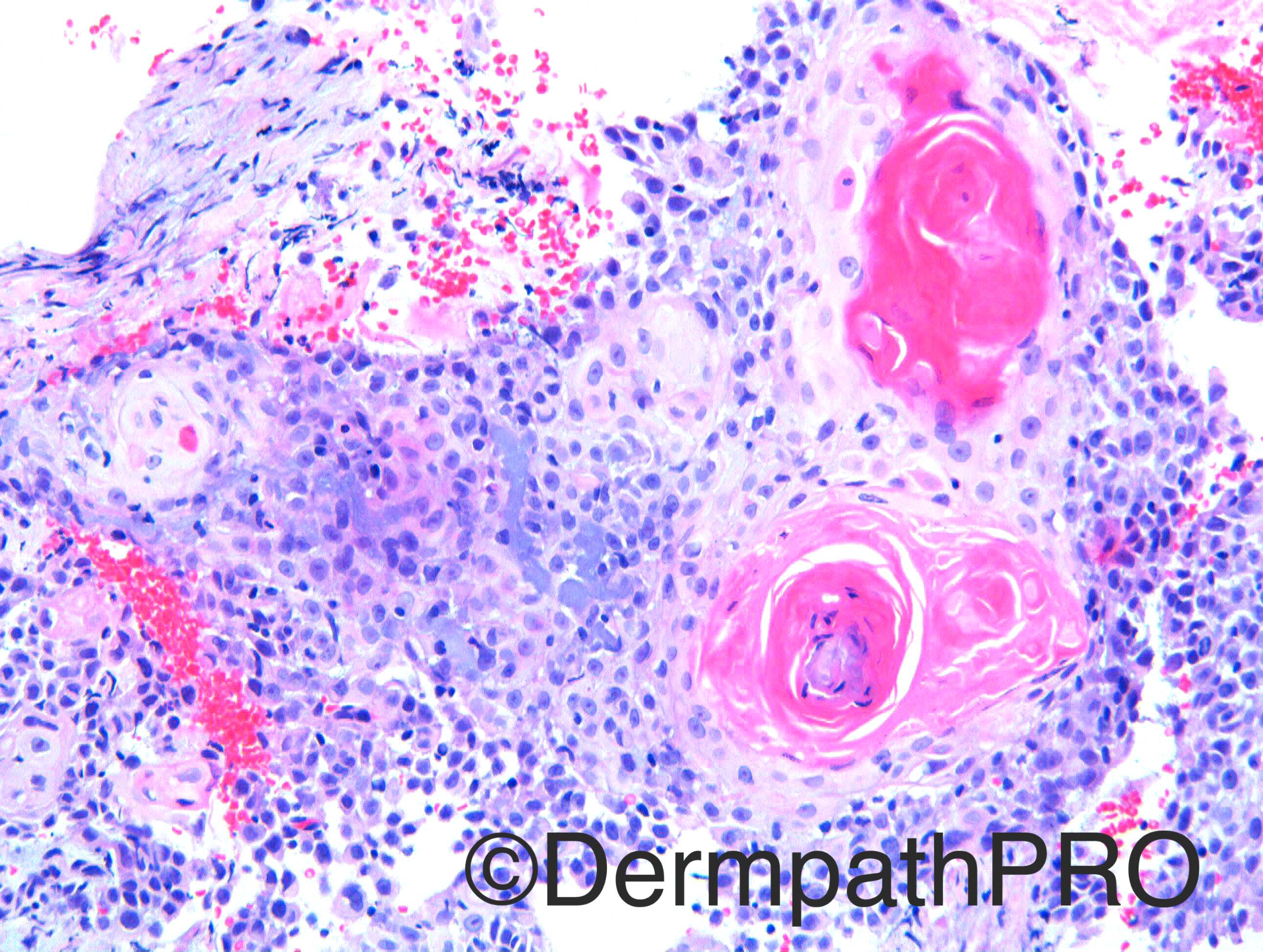
Case History: M90. ?SCC vertex of scalp

Case posted by Dr Richard Carr


  • Like 1
  Report Record

User Feedback


Nitin Khirwadkar

Posted

Agree with Mona. Invasive follicular SCC. Lovely mucin!

Share this comment


Link to comment
share_externally

Arti Bakshi

Posted

Yes, first thought is invasive SCC. However, the mucin is stromal rather than epithelial (the latter being a feature of Follicular SCC), so not sure about the significance.

Wondered if image 6 shows perforation of epithelium by elatic fibres?? Also some neutrophilic microabscesses. If this a rapidy growing lesion, perhaps proliferating phase of Keratoacanthoma comes in the d/d (although always difficult one to make on a small bx)

Share this comment


Link to comment
share_externally

Raul Perret

Posted

It seems that I arrived late this time. Agree with folicular SCC fits with the Warwick criteria. Just as a remark the lesión doesnt seem perfectly well circumscribed and it infiltrates with small cords of neoplastic cells, I imagine this is probable a more aggresive lesion than the ones that are perfectly well circumscribed with pushing borders...

Share this comment


Link to comment
share_externally

Raul Perret

Posted

Good remark Arti, I had the doubt with picture 6 too but I finally thought it was mucin, maybe I am wrong. Dr. Carr will illuminate us

Share this comment


Link to comment
share_externally

Raul Perret

Posted

Actually you are right, they are elastic fibers and the architecture is not the classical for follicular SCC, this fits better with proliferating KA, good job.

Share this comment


Link to comment
share_externally

Nitin Khirwadkar

Posted

Hmm, yes there is stromal mucin (usually not a feature of typical follicular SCCs), but there is some intra-tumoral mucin as well. Occasional micro-abscesses also present, along with perforation of elastic fibres, features of a KA! The cytological atypia in the last two images is a bit more for KA. Difficult case!

Share this comment


Link to comment
share_externally

vincenzo polizzi

Posted

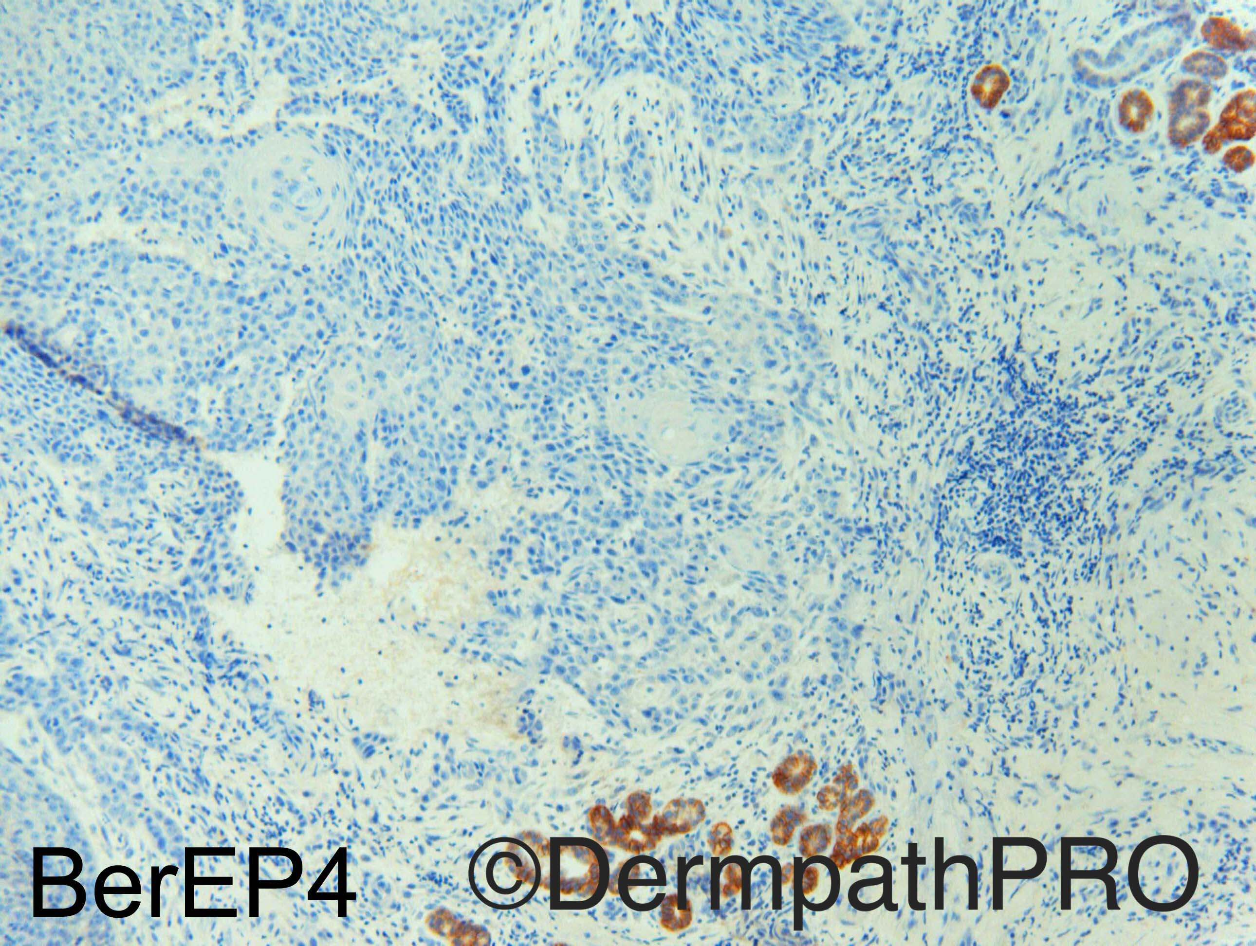
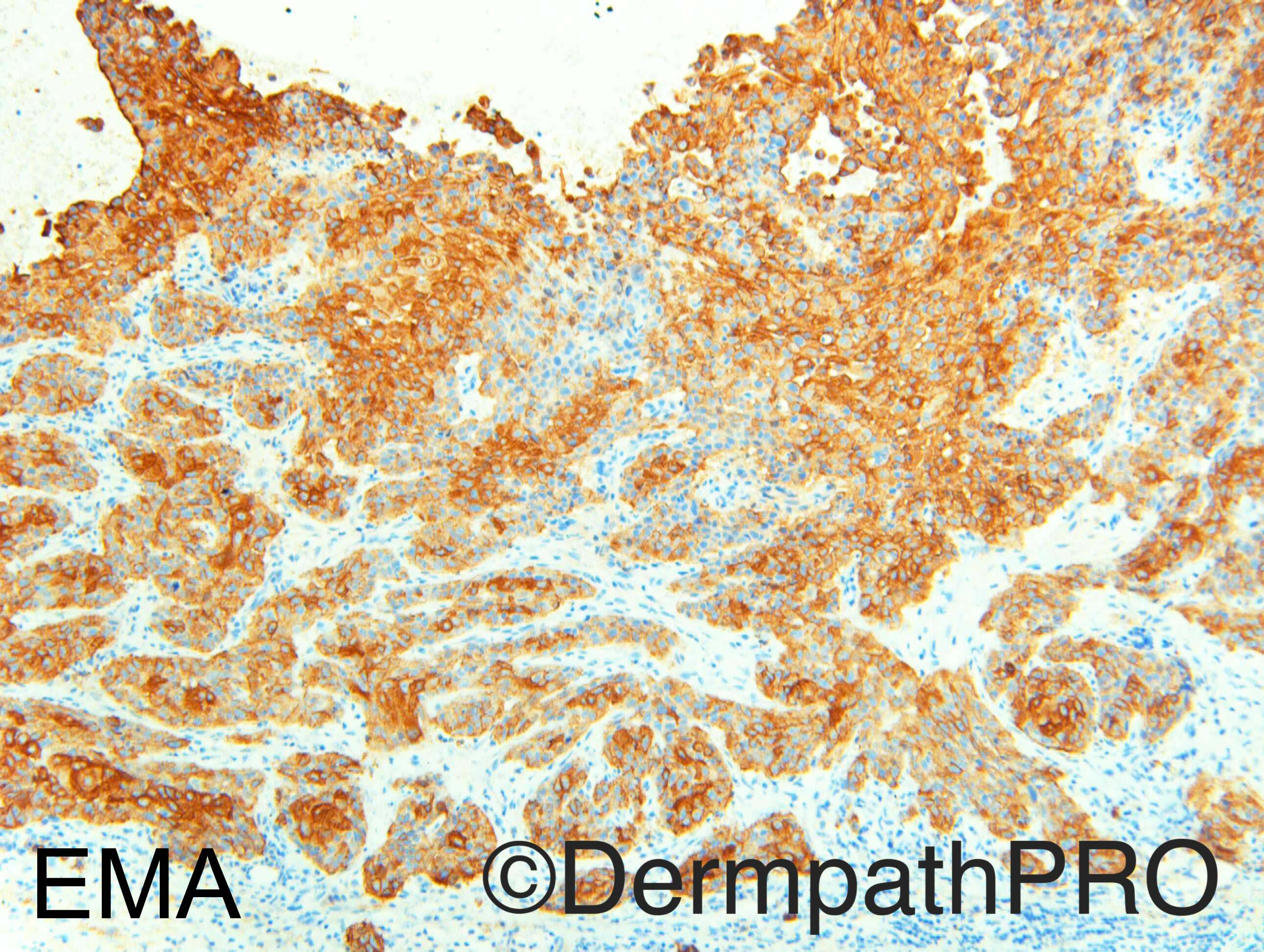
Immunostains suggest MAC, but morphologically i would had favoured DT...Difficult for me! I'm waiting for the diagnosis. 

Share this comment


Link to comment
share_externally

vincenzo polizzi

Posted

"Wondered if image 6 shows perforation of epithelium by elatic fibres?? Also some neutrophilic microabscesses. If this a rapidy growing lesion, perhaps proliferating phase of Keratoacanthoma" ...i agree...but it's too difficult for me seeing silhouette and other features of KA.

Share this comment


Link to comment
share_externally

Dr. Richard Carr

Posted

I called this a poorly differentiated squamous cell carcinoma (it may be follicular - but difficult to tell in this curettage).  The interstitial-type mucin is very rare in tumours other than BCC. I think I have only seen it once or twice previously in SCC, but I thought it made a nice contrast to last weeks case.  Immunostains are commensurate with SCC (negative BerEP4 in the basaloid cells and positive EMA even in the more basaloid cells, no glandular / luminal staining).  You are all right the mucin in follicular SCC is intra-epithelial and a useful clue to think of that diagnosis.  So far I have not observed intra-epithelial or extra-epithelial mucin in KA although intra-epithelial mucin it is not uncommonly seen in tricholemmoma, inverted follicular keratosis / seborrhoeic keratosis. There is elastic entrapment - if you check our review paper we reported that mainly in poorly differentiated subtypes of follicular SCC. Finding elastic entrapment in the mature central areas in KA is as far as I am currently aware fairly specific to KA (or infundibular psuedoepitheliomatous hyperplasia).  In this case the elastic entrapment is not in central matured areas but in the more solid basaloid infiltrative areas.  In KA we like to see full maturation (to cells with more abundant glassy cytoplasm than here) in the centre of all islands - in this biopsy I don't perceive that crucial feature.  There also appears to be foci of "spontaneous" acantholysis (i.e. not confined to neutrophil microabscess) a feature that I don't usually accept in KA.  Obviously if you are not sure you have to admit the uncertainty.  Be very wary of the tip of the iceberg effect in small biopsies (KA-like and craterifrom SCC that are superficially well differentiated) and remember KA is a clinicopathological team diagnosis in my view with many potential pitfalls. Clinically this case was not at all KA-like.  In fact this was a curettage of a crusted lesion on a patient with widespread severe actinic damage and many previously treated superficial lesions.  The lesion regrew into a 12mm nodule over the next 3 months and was finally excised with a full thickness skin graft and even then the deep margin was involved.

Share this comment


Link to comment
share_externally


Join the conversation

You can post now and register later. If you have an account, sign in now to post with your account.

Guest
Add a comment...

×   Pasted as rich text.   Paste as plain text instead

  Only 75 emoji are allowed.

×   Your link has been automatically embedded.   Display as a link instead

×   Your previous content has been restored.   Clear editor

×   You cannot paste images directly. Upload or insert images from URL.

Loading...

×
×
  • Create New...