Jump to content
In this section we have spot diagnoses posted on a daily basis since June 2010, now over 4000! You can review the archived cases and read the suggested diagnoses by users and the final comment by the contributors.
Case are uploaded each week day by 10 am UK time with the correct diagnosis will generally be posted at 8 pm UK time. Why not view the most recent spot diagnosis and proffer a diagnosis?

Case Number : Case 1530 - 05 May Posted By: Guest

Please read the clinical history and view the images by clicking on them before you proffer your diagnosis.
Submitted Date :
   (0 reviews)

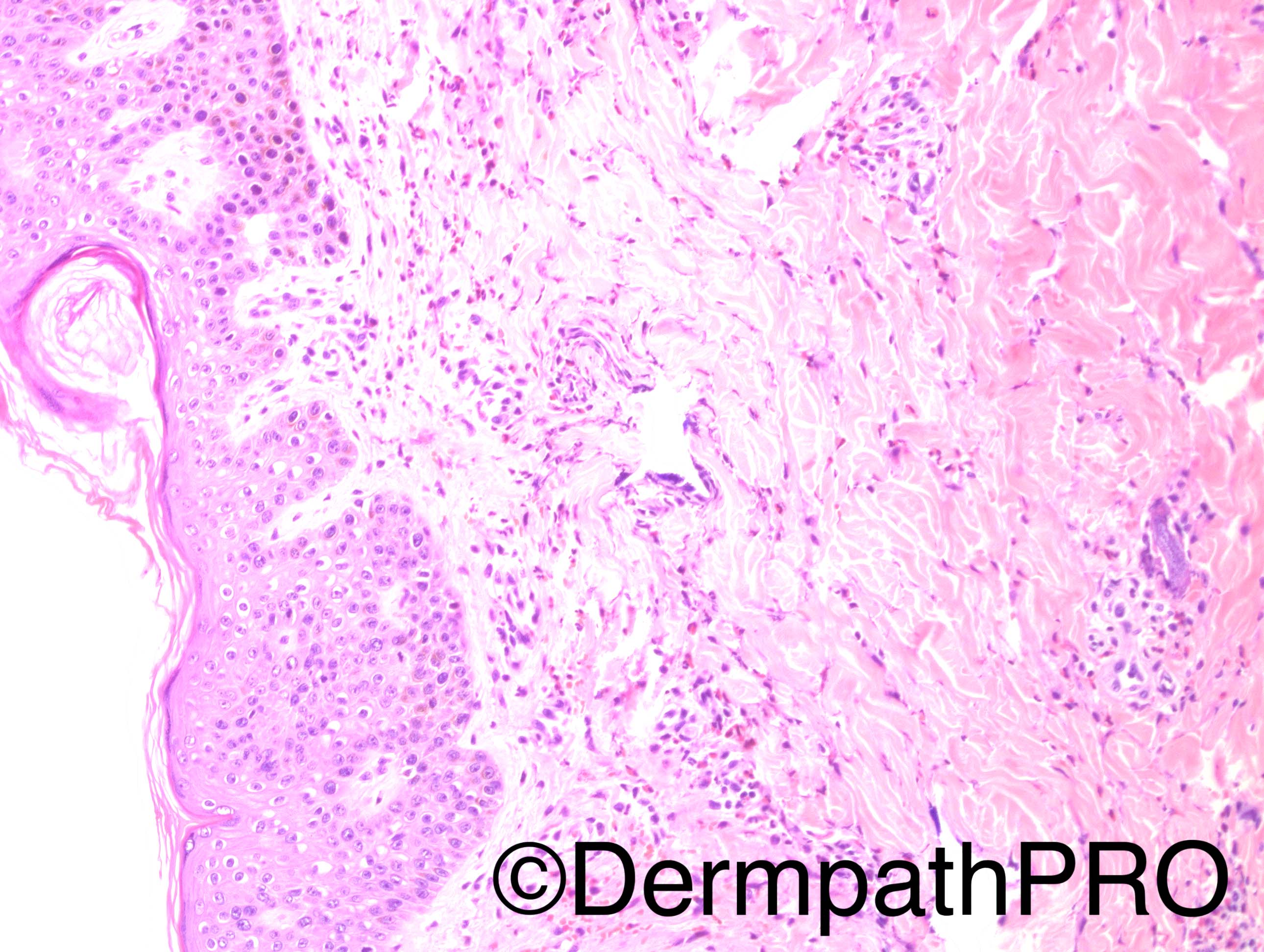
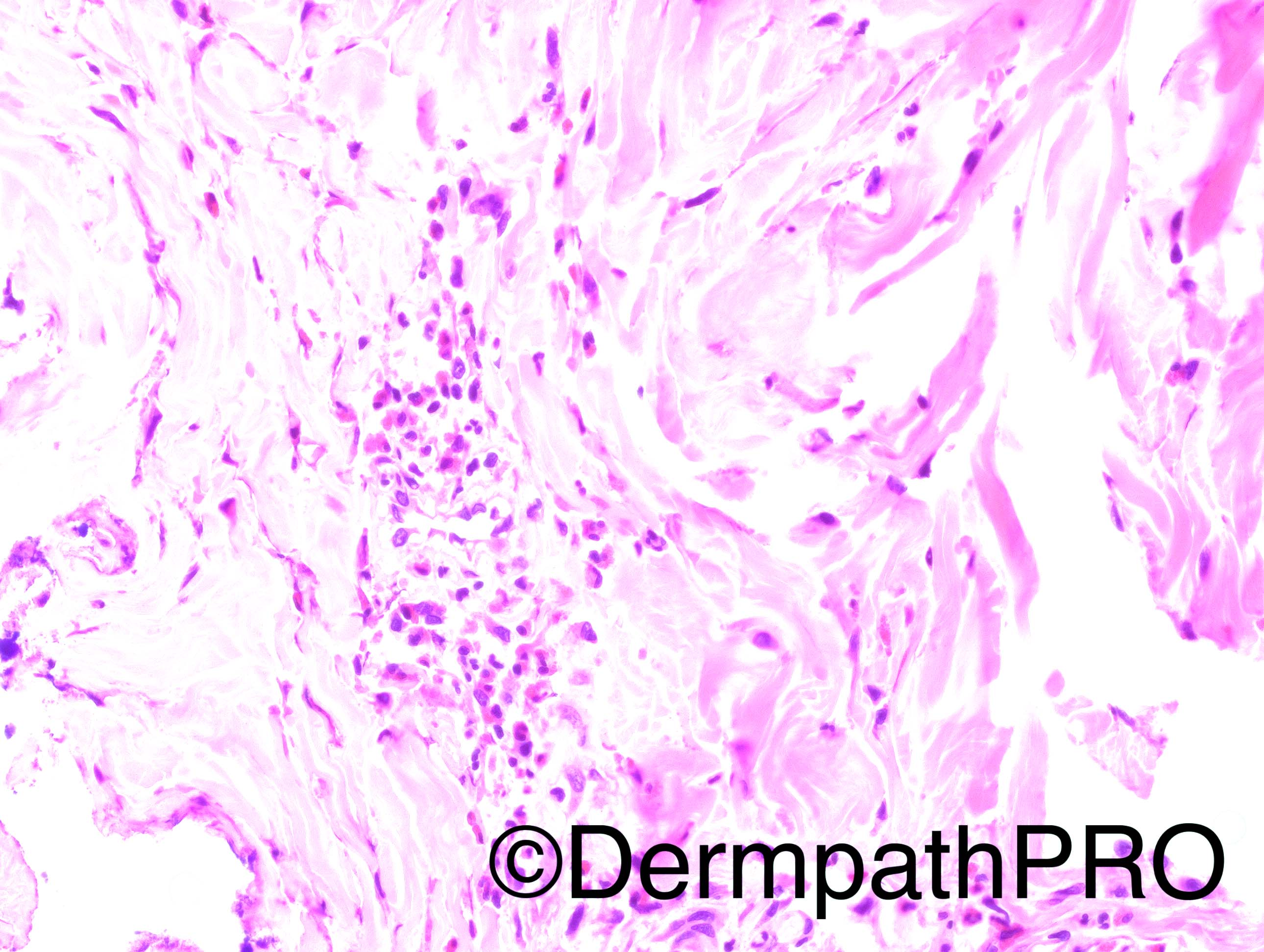
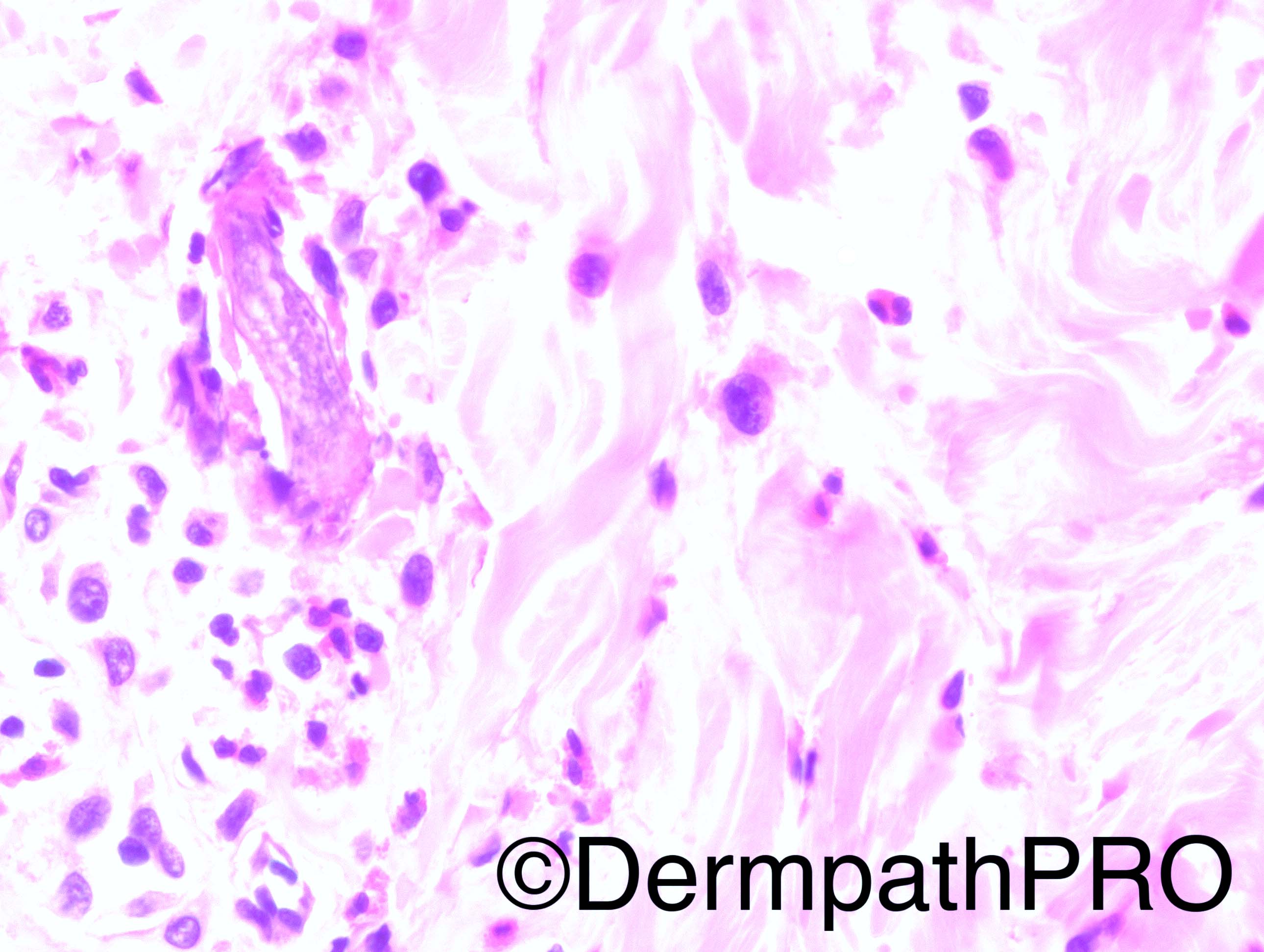
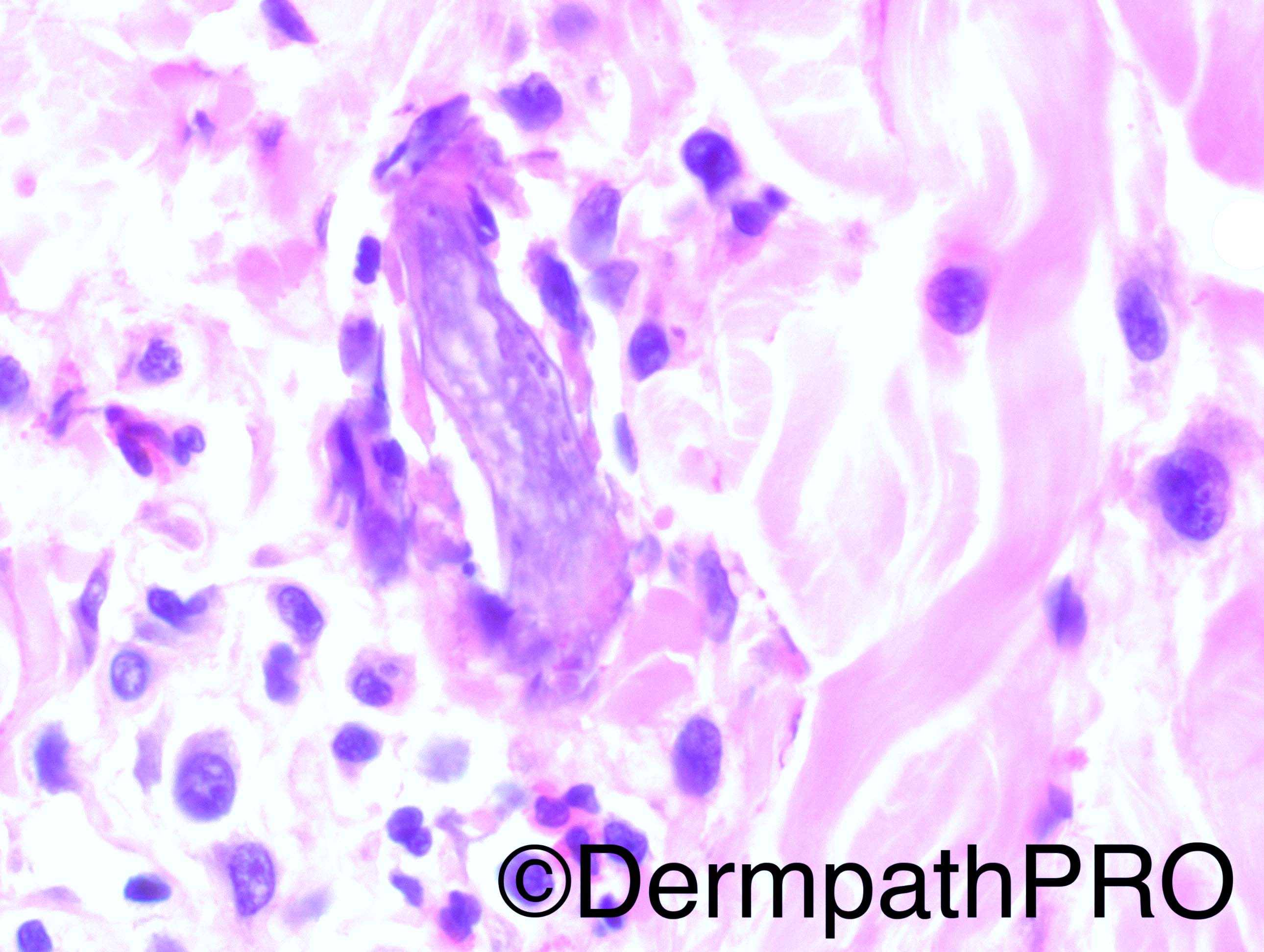
45 year-old Hispanic male with HIV and a CD4 count of 2, with red macules/rash. He is suspected to have TB meningitis. This biopsy is from an unspecified site.

Dr Hafeez Diwan


  Report Record

User Feedback


Guest Arash Daryakarr

Posted

I m thinking of well's syndrome. Not sure if this is a flame figure or some kind of parasite(e.g. microfilariae ?).

Share this comment


Link to comment
share_externally

IgorSC

Posted

It is a difficult case. I think this might be a cyst containing bradyzoites of Toxoplasmosis in the dermis.

Share this comment


Link to comment
share_externally

Raul Perret

Posted

I find it hard too, for me is too focalised, big and irregular for toxoplasmosis cyst. I was considering in the differential even an arthropod bite with rests persisting on the dermis (sting, etc)

Share this comment


Link to comment
share_externally

vincenzo polizzi

Posted

What about a eosinophilic folliculitis around a residual pilar shaft?

Share this comment


Link to comment
share_externally

Raul Perret

Posted

I watched with more attention and I agree with toxoplasmosis as you guys proposed

Share this comment


Link to comment
share_externally

Dr. Richard Carr

Posted

Strongyloidiasis to DDx.

Share this comment


Link to comment
share_externally

vincenzo polizzi

Posted

"What about a eosinophilic folliculitis around a residual pilar shaft?"-Demodex?

Share this comment


Link to comment
share_externally

Nitin Khirwadkar

Posted

Looks like a parasite, favour Strongyloidiasis.

Share this comment


Link to comment
share_externally

Dr. Hafeez Diwan

Posted

Strongyloidiasis.  This patient has a strongyloides hyperinfection (also seen in the CSF).

Share this comment


Link to comment
share_externally


Join the conversation

You can post now and register later. If you have an account, sign in now to post with your account.

Guest
Add a comment...

×   Pasted as rich text.   Paste as plain text instead

  Only 75 emoji are allowed.

×   Your link has been automatically embedded.   Display as a link instead

×   Your previous content has been restored.   Clear editor

×   You cannot paste images directly. Upload or insert images from URL.

Loading...

×
×
  • Create New...