Jump to content
In this section we have spot diagnoses posted on a daily basis since June 2010, now over 4000! You can review the archived cases and read the suggested diagnoses by users and the final comment by the contributors.
Case are uploaded each week day by 10 am UK time with the correct diagnosis will generally be posted at 8 pm UK time. Why not view the most recent spot diagnosis and proffer a diagnosis?

Case Number : Case 995 - 16th April Posted By: Guest

Please read the clinical history and view the images by clicking on them before you proffer your diagnosis.
Submitted Date :
   (0 reviews)

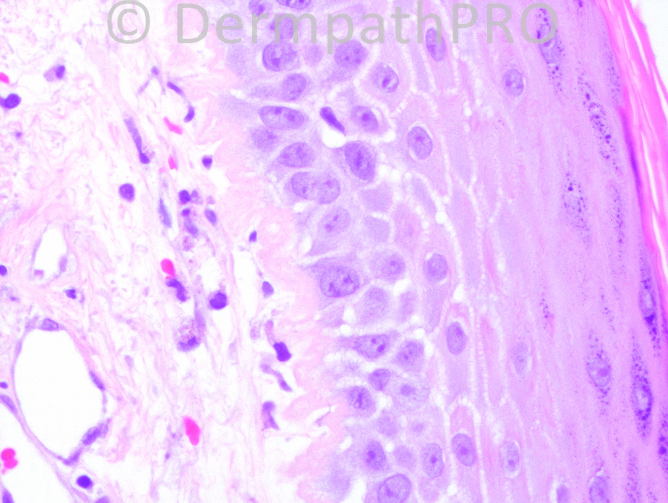
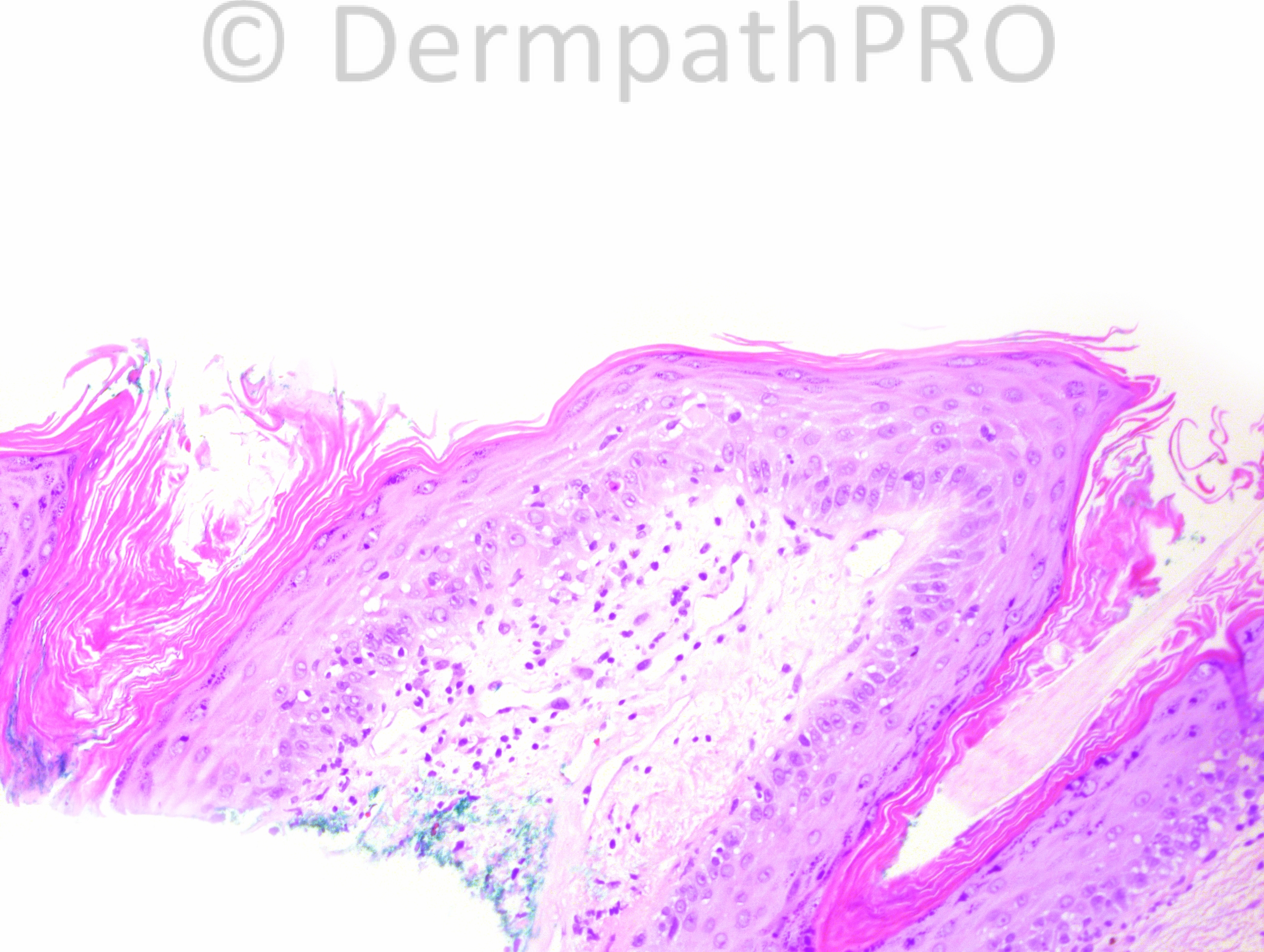
58 year-old female with left temporal scalp lesion.

Case posted by Dr. Hafeez Diwan.


  Report Record

User Feedback


Guest Tiberiu Tebeica

Posted

discoid lupus erythematosus vs. dermatomyositis

Share this comment


Link to comment
share_externally

Robledo F. Rocha

Posted

Epidermal atrophy with vacuolar change of the basal layer, follicular plugging, basement membrane thickening and dermal mucin deposition are signs of lupus erythematosus. But what about the spiking surface and the coarse keratohyaline granules? I think this a lesion of lupus erythematosus occurring in a site of a verruca vulgaris.

Share this comment


Link to comment
share_externally

Hanan Vaknine MD

Posted

[b]
Hypertrophic lupus erythematosus[/b]

Share this comment


Link to comment
share_externally

Dr. Hafeez Diwan

Posted

Discoid lupus erythematosus.

Share this comment


Link to comment
share_externally


Join the conversation

You can post now and register later. If you have an account, sign in now to post with your account.

Guest
Add a comment...

×   Pasted as rich text.   Paste as plain text instead

  Only 75 emoji are allowed.

×   Your link has been automatically embedded.   Display as a link instead

×   Your previous content has been restored.   Clear editor

×   You cannot paste images directly. Upload or insert images from URL.

Loading...

×
×
  • Create New...