Jump to content
In this section we have spot diagnoses posted on a daily basis since June 2010, now over 4000! You can review the archived cases and read the suggested diagnoses by users and the final comment by the contributors.
Case are uploaded each week day by 10 am UK time with the correct diagnosis will generally be posted at 8 pm UK time. Why not view the most recent spot diagnosis and proffer a diagnosis?

Case Number : Case 1102 - 12th September Posted By: Guest

Please read the clinical history and view the images by clicking on them before you proffer your diagnosis.
Submitted Date :
   (0 reviews)

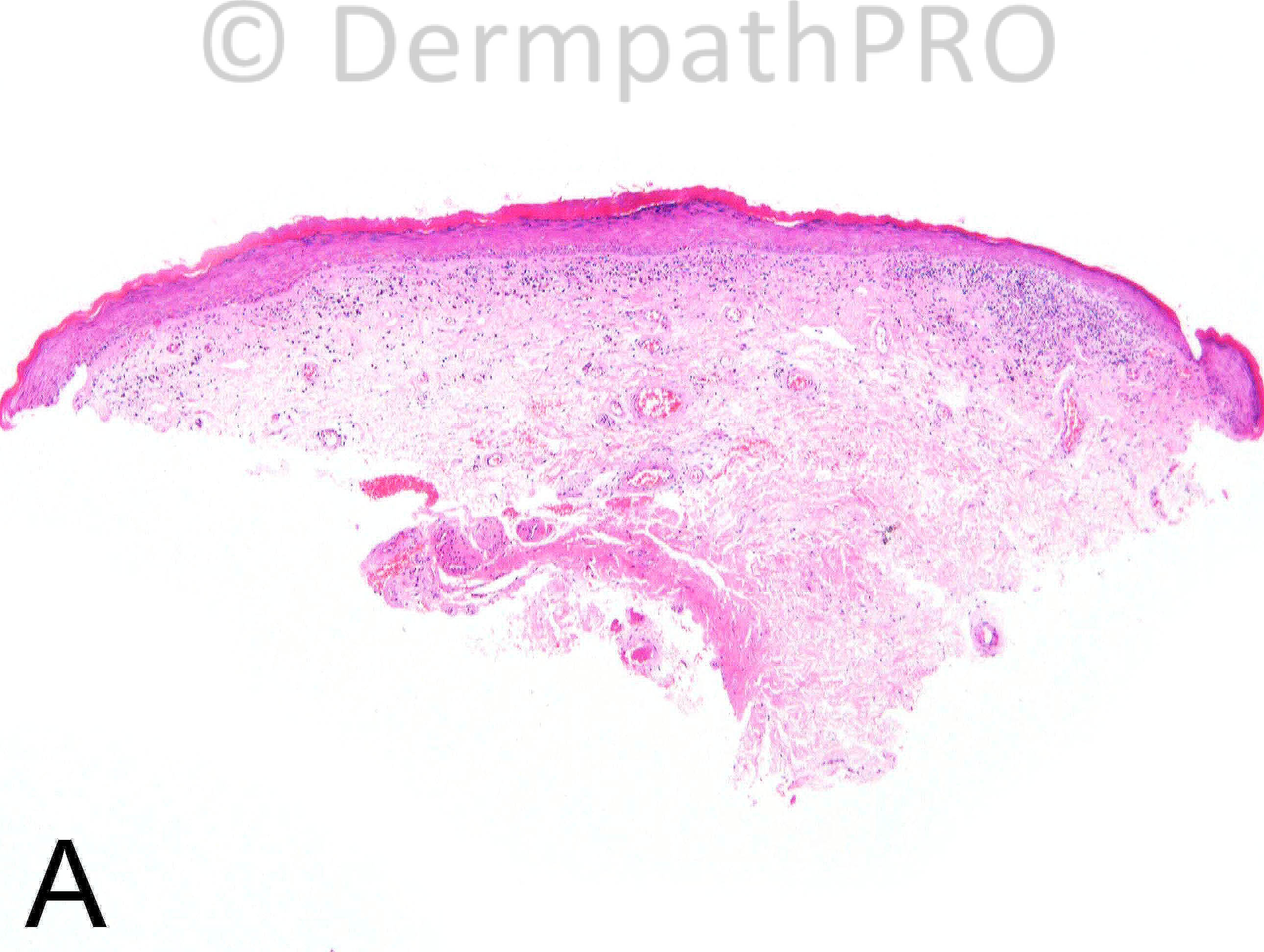
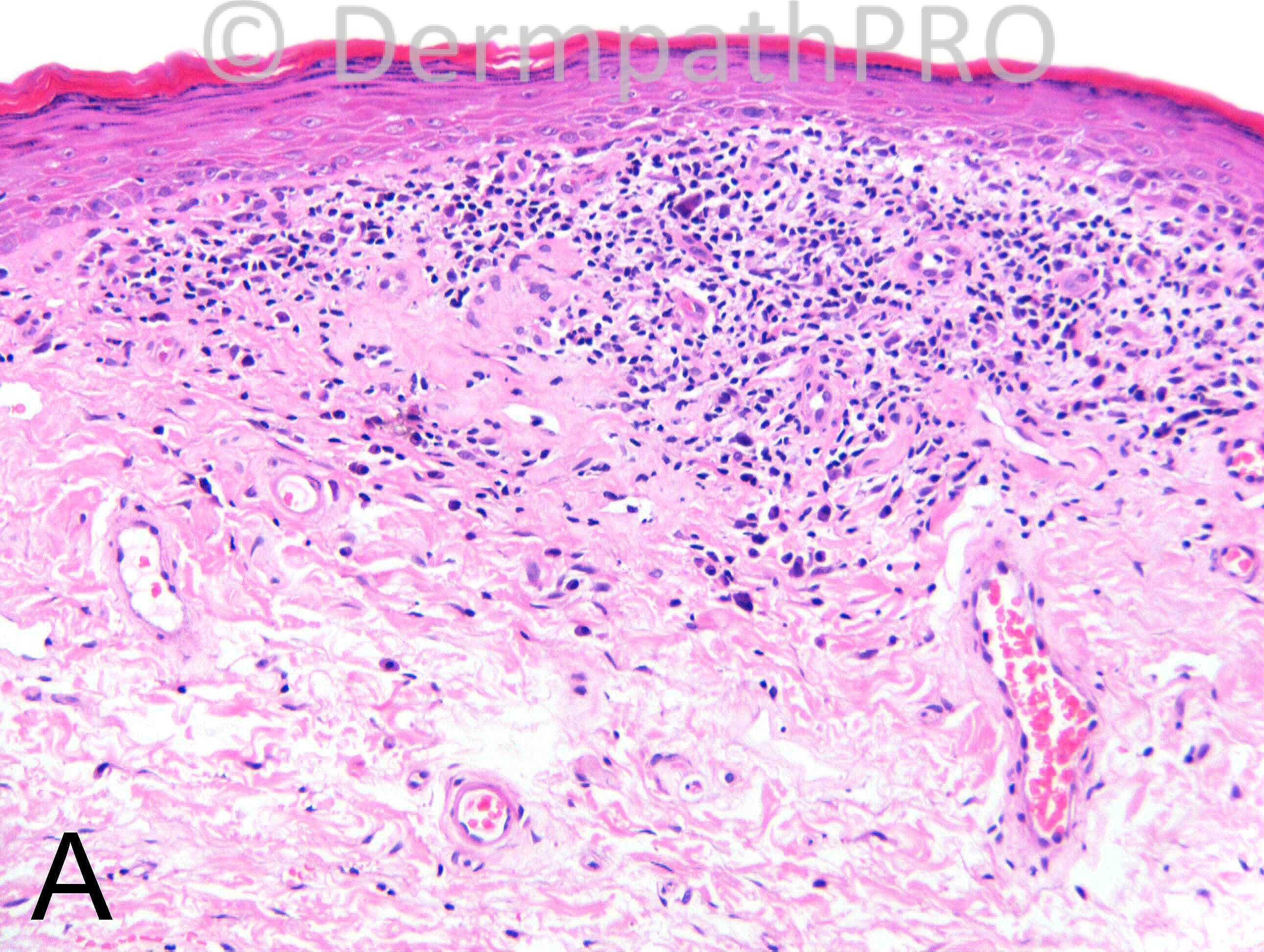
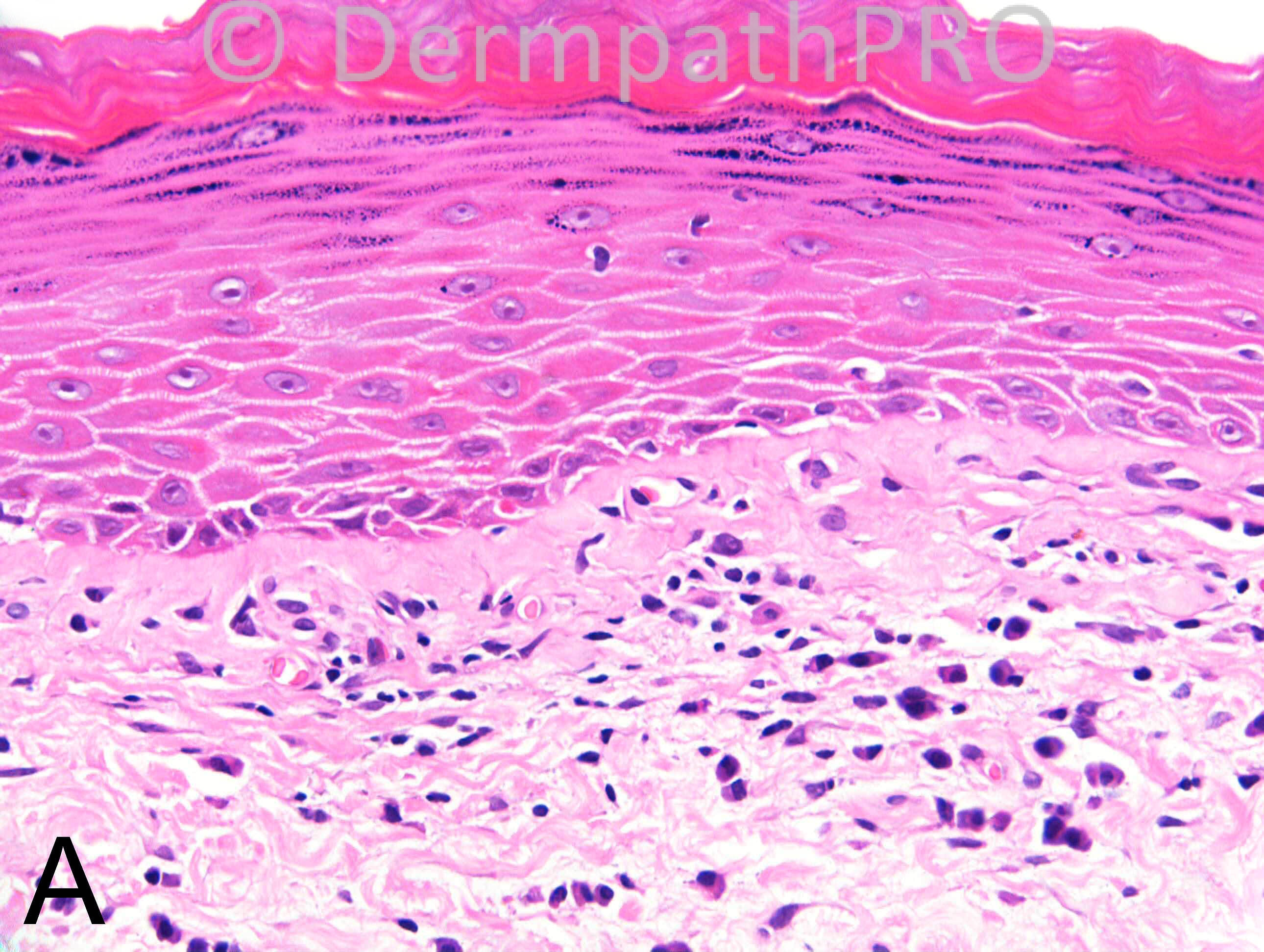
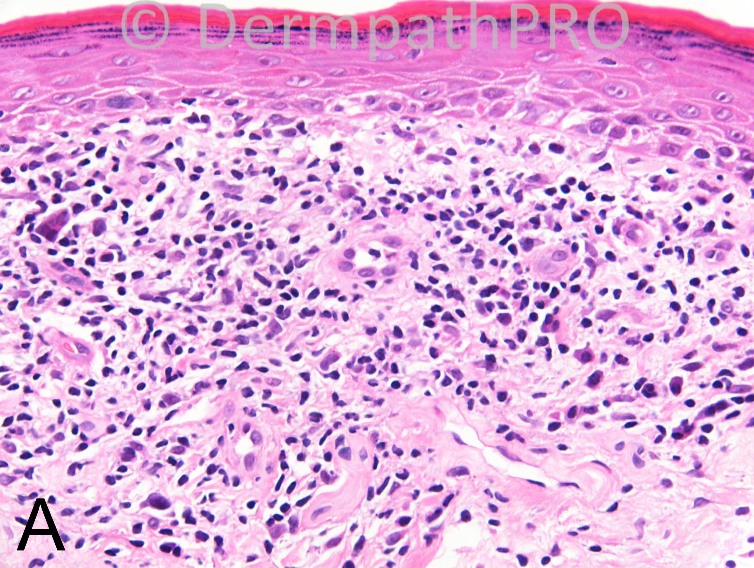
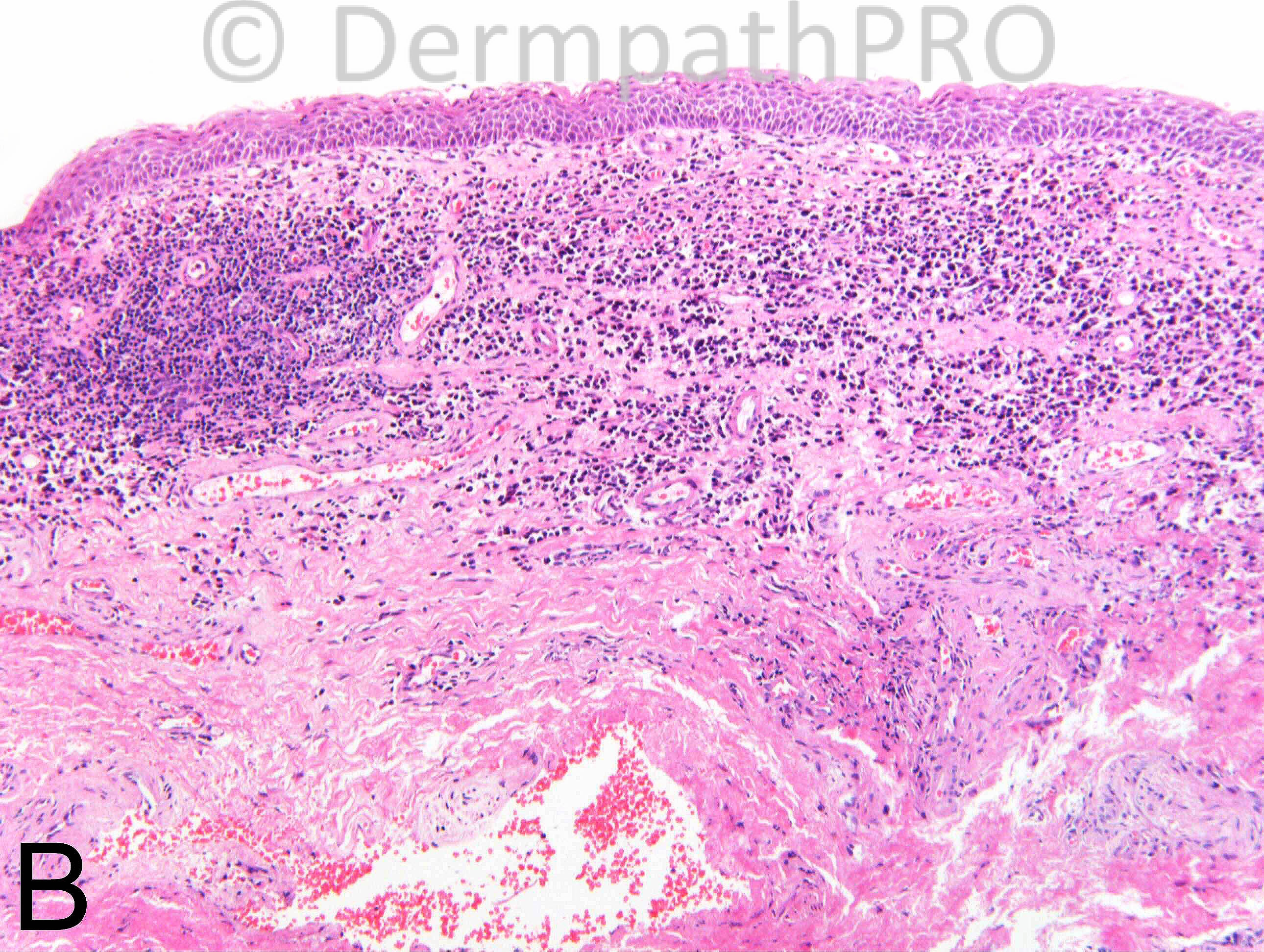
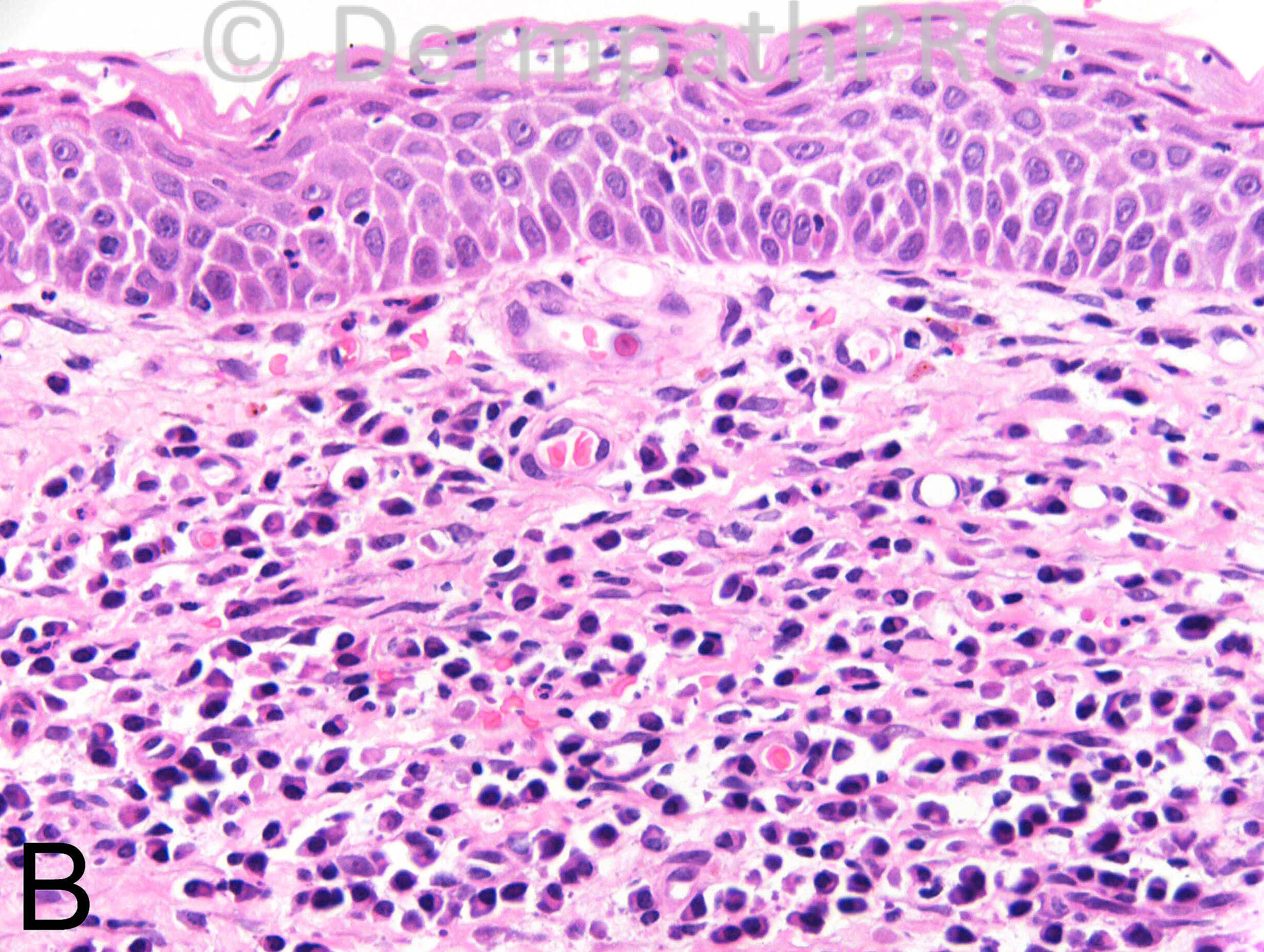
M50 4 years, well defined pale area (A) on glans penis with central ulcer (B).

Case Posted by Dr Richard Carr


  Report Record

User Feedback


Guest Romualdo

Posted

Zoon's balanitis pattern in the last two images and lichen sclerosus in the others.

Share this comment


Link to comment
share_externally

Inflammatory (presclerotic) lichen sclerosus. I don't think Zoon balanitis, which is [i]red[/i] in colour, fits with the clinical picture.

Share this comment


Link to comment
share_externally

Dr. Mona Abdel-Halim

Posted

I am looking at overlapping features of lichen sclerosus (early lichenoid phase) with features of Zoon's balanitis. Haven't found any reports of such association.

Share this comment


Link to comment
share_externally

Early lichen sclerosus. The clinical history and the atrophic epidermis along with hyperkeratosis are good tips.

Share this comment


Link to comment
share_externally

Guest Romualdo

Posted

Although my first comment was Zoon's balanitis I remembered a case presented by Dr. Richard Carr some time ago. According to hím , in almost all cases, what looks like Zoon's balanitis is in fact lichen sclerosus or lichen planus. In other words, it is probable that this diagosis will disappear in the future. I think this is lichen sclerosus with Zoon's balanitis pattern in some areas.

Share this comment


Link to comment
share_externally

Arti Bakshi

Posted

Early lichen sclerosus, agree with Romualdo's comment about Zoons balanitis like pattern.

Share this comment


Link to comment
share_externally

Dr. Richard Carr

Posted

Yes Romualdo am so glad you remembered that comment. I do believe the current thinking is that Zoon's is just a reaction pattern and indicates the patient has an underlying (invariably lichenoid) dermatosis but that the clinician has just biopsied the wrong bit for diagnosis. I have to admit that is how I tend to report Zoon's now (i.e. as a reaction pattern) and always suggest referring to a dermatologist (preferably with interest in vulval or penile disease) when the biopsy comes from a non-dermatologist. This is one of the few cases of Zoon's in which I have received a second biopsy showing the lichenoid dermatosis as I suspect clinicians are usually very reluctant to do multiple biopsies in this anatomic location! I actually erred towards lichen planus (just looks like the oral lichen planus we see all the time) but the essential point is to have the patient seen by a dermatologist who can correlate and provide the appropriate treatment. One point I would like to make is that mucosal lichen planus does not necessarily show saw-toothing or such prominent colloid bodies, may have plasma cells, and also can be more confluent and band-like although I appreciate LS is far more common generally. This case does show a hint of wedge-shaped hypergranulosis and I thought the clinical decription of a localised area may fit better too with LP. In females lichen planus can affect the inner aspects of the vulva / introitus whereas in LS the disease tends to spare the vagina / inner mucosal surface of the vulva. So the main message is look for the underlying disease when you see the Zoonoid pattern.
I hope you enjoy your weekends.

Share this comment


Link to comment
share_externally

Robledo F. Rocha

Posted

Papillary dermis is fibrotic and shows a sparse lichenoid infiltrate. Epidermis is atrophic and shows subtle wedge-shaped hypergranulosis and squamotization of the basal layer. So, I think it is lichen planus which, in mucosal sites, may present with erosive lesions and with infiltrate of plasma cells.

Share this comment


Link to comment
share_externally


Join the conversation

You can post now and register later. If you have an account, sign in now to post with your account.

Guest
Add a comment...

×   Pasted as rich text.   Paste as plain text instead

  Only 75 emoji are allowed.

×   Your link has been automatically embedded.   Display as a link instead

×   Your previous content has been restored.   Clear editor

×   You cannot paste images directly. Upload or insert images from URL.

Loading...

×
×
  • Create New...